

Memory encoding and dopamine in the aging brain: a psychopharmacological neuroimaging study
- PMID: 19625385
- PMCID: PMC2820708
- DOI: 10.1093/cercor/bhp139
Memory encoding and dopamine in the aging brain: a psychopharmacological neuroimaging study
Abstract
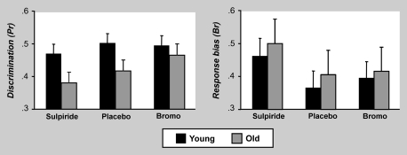
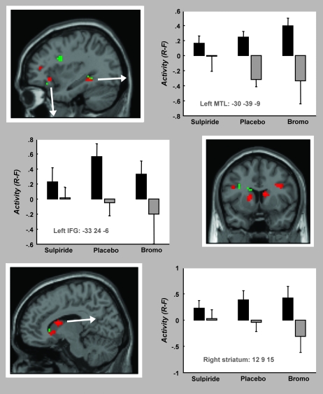
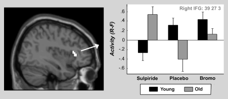
Normal aging brings with it changes in dopaminergic and memory functions. However, little is known about how these 2 changes are related. In this study, we identify a link between dopamine, episodic memory networks, and aging, using pharmacological functional magnetic resonance imaging. Young and older adults received a D2-like agonist (Bromocriptine, 1.25 mg), a D2-like antagonist (Sulpiride, 400 mg), and Placebo, in a double-blind crossover procedure. We observed group differences, during memory encoding, in medial temporal, frontal, and striatal regions and moreover, these regions were differentially sensitive across groups to dopaminergic perturbation. These findings suggest that brain systems underlying memory show age-related changes and that dopaminergic function may be key in understanding these changes. That these changes have behavioral consequences was suggested by the observation that drug modulations were most pronounced in older subjects with poorer recognition memory. Our findings provide direct evidence linking ageing, memory, and dopaminergic change.
Figures

Similar articles
-
A functional MRI study of the effects of bromocriptine, a dopamine receptor agonist, on component processes of working memory.Psychopharmacology (Berl). 2005 Aug;180(4):644-53. doi: 10.1007/s00213-005-0077-5. Epub 2005 Sep 14. Psychopharmacology (Berl). 2005. PMID: 16001111 Clinical Trial.
-
Simulating neurocognitive aging: effects of a dopaminergic antagonist on brain activity during working memory.Biol Psychiatry. 2010 Mar 15;67(6):575-80. doi: 10.1016/j.biopsych.2009.12.013. Biol Psychiatry. 2010. PMID: 20138255
-
Dopamine and memory dedifferentiation in aging.Neuroimage. 2017 Jun;153:211-220. doi: 10.1016/j.neuroimage.2015.03.031. Epub 2015 Mar 21. Neuroimage. 2017. PMID: 25800211 Free PMC article. Clinical Trial.
-
Dopaminergic challenge with bromocriptine one month after mild traumatic brain injury: altered working memory and BOLD response.J Neuropsychiatry Clin Neurosci. 2011 Summer;23(3):277-86. doi: 10.1176/jnp.23.3.jnp277. J Neuropsychiatry Clin Neurosci. 2011. PMID: 21948888 Free PMC article. Clinical Trial.
-
Age differences in the neural correlates of novelty processing: The effects of item-relatedness.Brain Res. 2015 Jul 1;1612:2-15. doi: 10.1016/j.brainres.2014.08.006. Epub 2014 Aug 19. Brain Res. 2015. PMID: 25149192 Review.
Cited by
-
Stress modulates reinforcement learning in younger and older adults.Psychol Aging. 2013 Mar;28(1):35-46. doi: 10.1037/a0029823. Epub 2012 Sep 3. Psychol Aging. 2013. PMID: 22946523 Free PMC article. Clinical Trial.
-
Vitamin K2 (Menaquinone-7) Reverses Age-Related Structural and Cognitive Deterioration in Naturally Aging Rats.Antioxidants (Basel). 2022 Mar 8;11(3):514. doi: 10.3390/antiox11030514. Antioxidants (Basel). 2022. PMID: 35326164 Free PMC article.
-
Aging and recognition memory: A meta-analysis.Psychol Bull. 2019 Apr;145(4):339-371. doi: 10.1037/bul0000185. Epub 2019 Jan 14. Psychol Bull. 2019. PMID: 30640498 Free PMC article.
-
Normal aging modulates prefrontoparietal networks underlying multiple memory processes.Eur J Neurosci. 2012 Dec;36(11):3559-67. doi: 10.1111/j.1460-9568.2012.08254.x. Epub 2012 Aug 21. Eur J Neurosci. 2012. PMID: 22909094 Free PMC article.
-
Dopamine modulates the neural representation of subjective value of food in hungry subjects.J Neurosci. 2014 Dec 10;34(50):16856-64. doi: 10.1523/JNEUROSCI.2051-14.2014. J Neurosci. 2014. PMID: 25505337 Free PMC article. Clinical Trial.
References
-
- Abrams R, Taylor MA. Cognitive dysfunction in melancholia. Psychol Med. 1987;17:359–362. - PubMed
-
- Addis DR, McAndrews MP. Prefrontal and hippocampal contributions to the generation and binding of semantic associations during successful encoding. Neuroimage. 2006;33(4):1194–1206. - PubMed
-
- Aizenstein HJ, Clark KA, Butters MA, Cochran J, Stenger VA, Meltzer CC, Reynolds CF, Carter CS. The BOLD hemodynamic response in healthy aging. J Cogn Neurosci. 2004;16(5):786–793. - PubMed
-
- Amenta F, Mignini F, Ricci A, Sabbatini M, Tomassoni D, Tayebati SK. Age-related changes of dopamine receptors in the rat hippocampus: a light microscope autoradiography study. Mech Ageing Dev. 2001;122(16):2071–2083. - PubMed
Publication types
MeSH terms
Substances
Grants and funding
LinkOut - more resources
Full Text Sources
Medical
