. 2009 Oct;83(19):10187-97.
doi: 10.1128/JVI.00396-09. Epub 2009 Jul 22.

Human cytomegalovirus UL28 and UL29 open reading frames encode a spliced mRNA and stimulate accumulation of immediate-early RNAs

Affiliations

Human cytomegalovirus UL28 and UL29 open reading frames encode a spliced mRNA and stimulate accumulation of immediate-early RNAs

Dora P Mitchell et al. J Virol. 2009 Oct.

Abstract

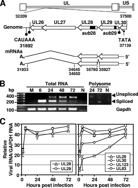
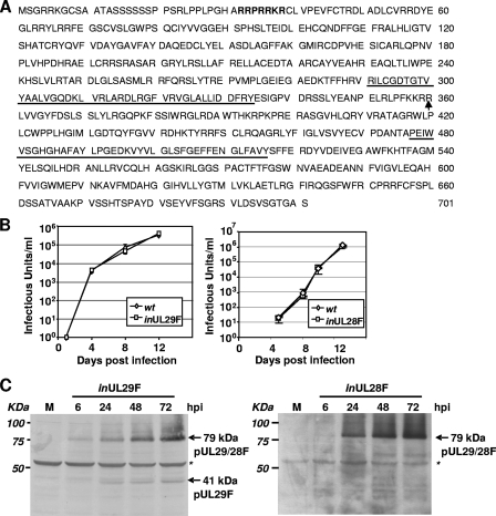
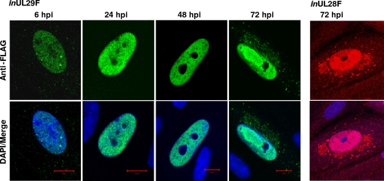
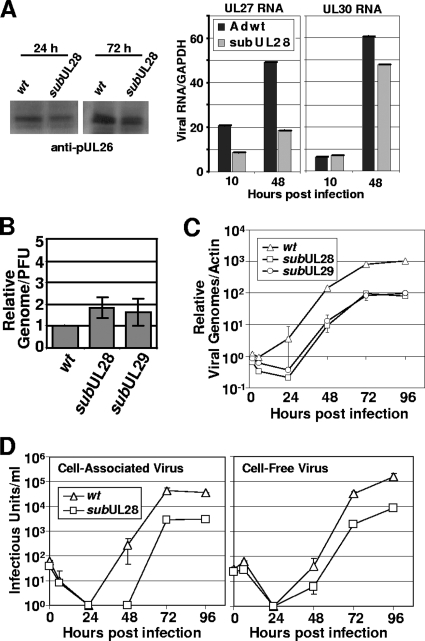
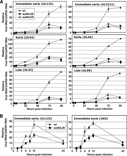
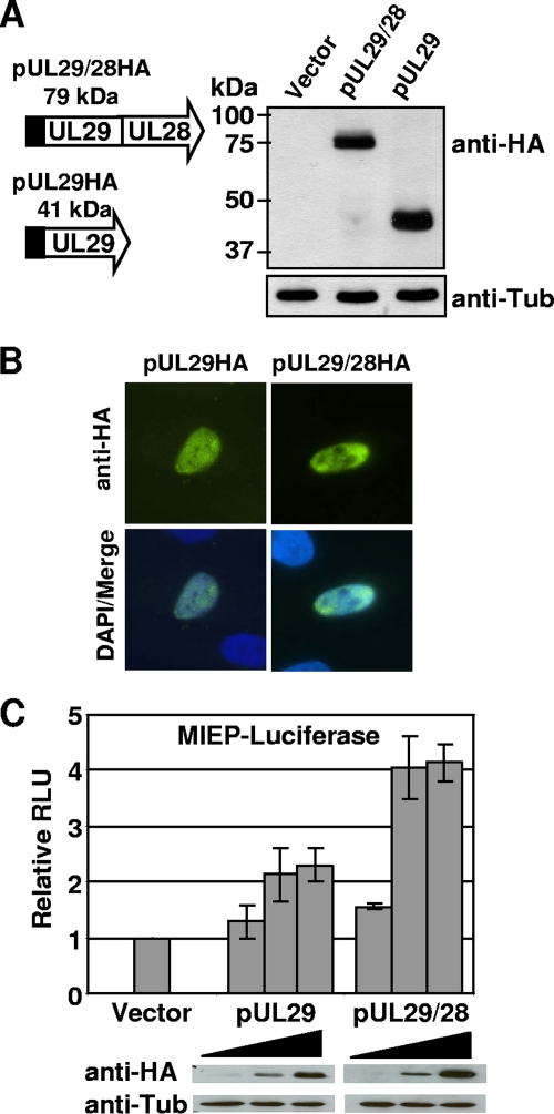
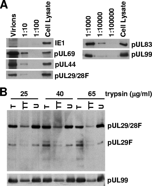
We have identified a spliced transcript that contains sequences from the HCMV UL29 and UL28 open reading frames. It contains amino-terminal UL29 sequences followed by UL28 sequences, and it includes a poly(A) signal derived from the 3'-untranslated region following the UL26 open reading frame. UL29/28 RNA is expressed with early kinetics, and a virus containing a FLAG epitope inserted at the amino terminus of UL29 expressed a tagged approximately 79-kDa protein, pUL29/28, that was detected at 6 h postinfection. The virus also expressed a less-abundant tagged 41-kDa protein, which corresponds in size to a protein that could be produced by translation of an unspliced UL29/28 transcript. Consistent with this prediction, both unspliced and spliced UL29/28 transcript was present in RNA isolated from polysomes. FLAG-tagged protein from the UL29/28 locus accumulated within nuclear viral replication centers during the early phase of infection. Late after infection it was present in the cytoplasm as well, and the protein was present and resistant to proteinase treatment in partially purified preparations of viral particles. Disruption of the UL29/28 locus by mutation resulted in a 10-fold decrease in the levels of DNA replication along with a similar reduction in virus yield. Quantitative reverse transcription-PCR analysis revealed an approximately 2-fold decrease in immediate-early gene expression at 4 to 10 h postinfection compared to the wild-type virus, and transient expression of pUL29/28 activated the major immediate-early promoter. Our results argue that the UL29/28 locus contributes to activation of immediate-early gene expression.


Figures

FIG. 1.
FIG. 1.
HCMV-coded transcripts containing the UL28 and UL29 genes. (A) Location of the UL28 and 29 ORFs on the viral genome and organization of their transcripts. The top of the panel diagrams the coding region showing the location of the UL26-UL30 ORFs within the viral chromosome. The coding sequences are indicated by open arrows with the C terminus of each ORF indicated by an arrowhead. Also indicated are the locations of three polyadenylation signals (filled circles) and a putative TATA box element (arrow). The position of deletions in substitution mutations within the UL28 and UL29 genes of the recombinant viruses, BADsubUL28 and BADsubUL29, are indicated by filled boxes. The bottom of the panel shows UL28 and UL29-related mRNAs with the location of splice donor and acceptor sites noted. Splice sites were identified by 3′ RACE analysis using RNA isolated at 6 and 24 hpi from fibroblasts infected with wild-type HCMV. The nucleotide numbers are from GenBank accession no. X17403. (B) Expression of unspliced and spliced transcripts during infection. Total RNA was isolated at indicated times from fibroblasts infected at a multiplicity of 0.5 infectious unit/cell with wild-type virus. RT-PCR was performed using primers that span a predicted splice donor at nt 35927 and an acceptor at nt 35780 and produced products corresponding to both spliced (188 nt) and unspliced (334 nt) cDNAs. Primers to GAPDH were used for a loading control. PCR products for both spliced and unspliced sequences were also observed using cDNA prepared from purified polysomes but not in the non-RT (N) control. (C) Accumulation of RNAs containing UL28 and UL29 sequences after infection with wild-type virus. Fibroblasts were infected under the conditions described above. Total RNA was isolated at the indicated times after infection, and RNA was quantified by real-time RT-PCR using primers specific to UL28 and UL29 and normalized to GAPDH RNA. RNAs containing the UL26, UL30, UL123, and UL83 ORFs were analyzed as controls for the rate of accumulation of viral RNAs from different kinetic classes.
FIG. 2.
FIG. 2.
Early expression and predominantly nuclear localization of HCMV pUL29/28. (A) Amino acid sequence of HCMV strain AD169 pUL29/28 encoded by the spliced transcript encoding the UL29 and UL28 genes. The location of the junction between UL29 and UL28 is indicated by an arrow. A putative nuclear localization signal is in boldface letters, and the US22 family domains found in both UL29 and UL28 are underlined. (B) Virus expressing epitope-tagged pUL29/28 grows with normal kinetics. Replicate cultures of fibroblasts were infected at a multiplicity of 0.5 infectious unit/cell with wild-type HCMV (wt) or a recombinant virus containing a FLAG epitope at the N terminus of the UL29 ORF (inUL29F) and at the C terminus of the UL28 ORF (inUL28F). Culture supernatants were harvested at the indicated times, and the infectious virus progeny was quantified. (C) Accumulation of tagged proteins in cells infected with inUL29F and inUL28F viruses. Fibroblasts were infected at a multiplicity of 3.0 PFU/cell, harvested at the indicated times, and processed for Western blot assay using a FLAG-specific antibody. A nonspecific band (asterisks) was monitored to confirm equal protein loading.
FIG. 3.
FIG. 3.
Localization of pUL29/28F in fibroblasts during infection. Cells were infected at a multiplicity of 0.5 infectious unit/cell using either inUL29F or inUL28F, fixed at the indicated times, and processed for immunofluorescence using a FLAG-specific antibody (inUL29F, green; inUL28F, red) and DAPI (blue).
FIG. 4.
FIG. 4.
Inefficient BADsubUL28 and BADsubUL29 replication. (A) Expression of UL26, UL27, and UL30 in fibroblasts infected with subUL28 compared to wild-type virus. Fibroblasts were infected using equivalent amounts of virus, harvested at the indicated times, and processed for Western blot analysis with a pUL26-specific antibody. Expression of RNAs containing UL27 and UL30 ORFs were quantified by qRT-PCR using total RNA harvested from cells infected by wild-type and subUL28 viruses. (B) Infectivity of wild-type, subUL28, and subUL29 viruses. Genome content was determined by using qPCR and primers against HCMV DNA using partially purified virus, and infectious units were determined by quantifying IE1-positive cells using the same virus stock. The results are from duplicate experiments and are presented relative to wild-type virus. (C) Accumulation of viral DNA. Fibroblasts were infected with wild type, subUL28, or subUL29 at an input genome number equivalent to 0.1 infectious unit of wild-type virus/cell. Total cell-associated DNA was isolated, and viral genomes were quantified by using real-time PCR and normalized to β-actin DNA. (D) Single-step growth analysis of subUL28. Cell-associated (left panel) and cell-free (right panel) virus was assayed. Fibroblasts were infected with wild-type or subUL28 virus at an input genome number equivalent to 0.1 infectious unit of wild-type virus/cell. Infected cell culture medium was collected as cell-free virus samples, and cell-associated virus was isolated by freezing and thawing cell pellets. The amount of virus present in each sample was determined by counting the number of IE1-positive cells and experiment was completed in duplicate. wt, wild type.
FIG. 5.
FIG. 5.
pUL29/28-deficient viruses express reduced levels of viral transcripts. (A) Accumulation of viral RNAs during a single-step growth analysis. Fibroblasts were infected with wild-type (wt), subUL28, or subUL29 at an input genome number equivalent to 0.1 infectious unit of wild-type virus/cell. Total RNA was isolated at the indicated times after infection, and viral RNA was quantified by real-time RT-PCR using primers specific to the indicated genes and normalized to GAPDH RNA. (B) Accumulation of HCMV RNAs during the immediate-early phase of infection. Total RNA was isolated at the indicated times postinfection, and RNA levels were quantified as described above using primers specific to the immediate-early genes UL123 and US3.
FIG. 6.
FIG. 6.
pUL29/28 activates the MIEP. (A) Transient expression of pUL29/28 and pUL29. The UL29/28 and UL29 sequences were introduced into mammalian expression vector pCGN in-frame with the HA tag. 293T cells were transfected using empty vector or the pUL29/28HA or pUL29HA expression vectors, and whole-cell lysates were analyzed by Western blotting using an antibody to HA or to cellular tubulin. (B) Localization of pUL29/28HA and pUL29HA was visualized by indirect immunofluorescence on transfected cells using anti-HA (green) and DAPI (blue). (C) 293T cells were transfected using 50 ng of pGL3-MIEP reporter plasmid and 10, 100, or 500 ng of pCGN empty vector, pCGN-pUL29/28, or pCGN-pUL29 effector plasmid. Luciferase activity was assayed 48 h posttransfection using equal protein amounts within each lysate and normalized to luciferase activity from empty vector. The levels of pUL29/28HA and pUL29HA expression were assayed by Western blot analysis using the same lysates and antibody to HA or cellular tubulin.
FIG. 7.
FIG. 7.
pUL29 and pUL29/28 are present in HCMV virions. (A) Partially purified BADinUL29F virion preparations contain pUL29/28F. Virions were solubilized, and serial dilutions, as well as an infected cell lysate control, were analyzed by Western blotting with antibodies to the FLAG epitope and the known virion proteins pUL69, pUL44, pUL83, and pUL99. pUL123 was assayed as a negative control. (B) pUL29F and pUL29/28F are located within the virion envelope. inUL29F virions were treated with increasing amounts of trypsin (T), with trypsin and Triton X-100 (TT), or untreated (U). Virions were then solubilized, and proteins were analyzed by Western blotting with a monoclonal antibody specific to the FLAG epitope and, as a control, a monoclonal antibody to pUL99.

Similar articles

Cited by

References

    1. Adamo, J. E., J. Schroer, and T. Shenk. 2004. Human cytomegalovirus TRS1 protein is required for efficient assembly of DNA-containing capsids. J. Virol. 78:10221-10229. - PMC - PubMed
    1. Baldick, C. J., Jr., A. Marchini, C. E. Patterson, and T. Shenk. 1997. Human cytomegalovirus tegument protein pp71 (ppUL82) enhances the infectivity of viral DNA and accelerates the infectious cycle. J. Virol. 71:4400-4408. - PMC - PubMed
    1. Blais, J. D., V. Filipenko, M. Bi, H. P. Harding, D. Ron, C. Koumenis, B. G. Wouters, and J. C. Bell. 2004. Activating transcription factor 4 is translationally regulated by hypoxic stress. Mol. Cell. Biol. 24:7469-7482. - PMC - PubMed
    1. Bowen, N. J., N. Fujita, M. Kajita, and P. A. Wade. 2004. Mi-2/NuRD: multiple complexes for many purposes. Biochim. Biophys. Acta 1677:52-57. - PubMed
    1. Bresnahan, W. A., and T. E. Shenk. 2000. UL82 virion protein activates expression of immediate-early viral genes in human cytomegalovirus-infected cells. Proc. Natl. Acad. Sci. USA 97:14506-14511. - PMC - PubMed

Publication types

LinkOut - more resources