

Matrix and envelope coevolution revealed in a patient monitored since primary infection with human immunodeficiency virus type 1
- PMID: 19625403
- PMCID: PMC2748007
- DOI: 10.1128/JVI.01213-09
Matrix and envelope coevolution revealed in a patient monitored since primary infection with human immunodeficiency virus type 1
Abstract
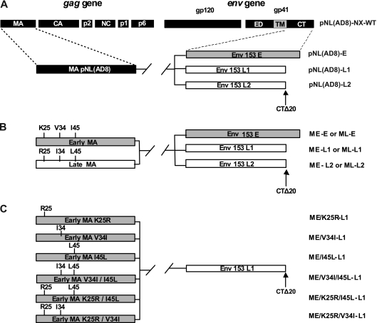
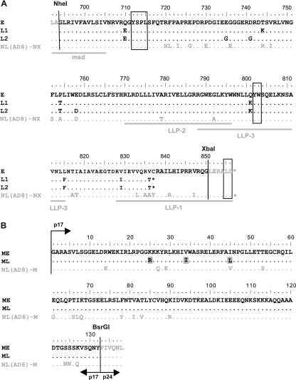
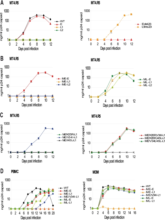
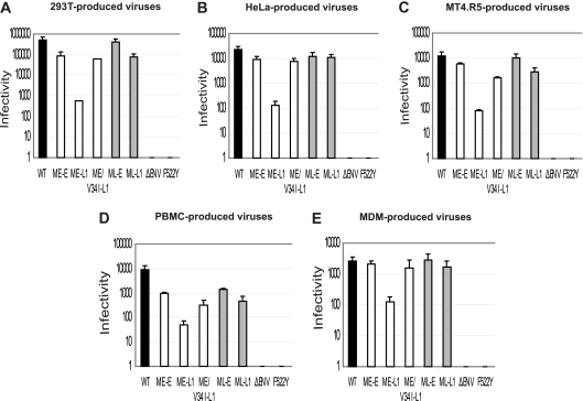
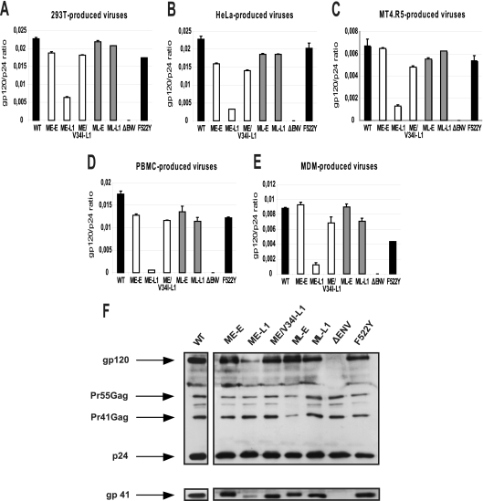
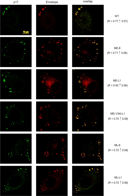
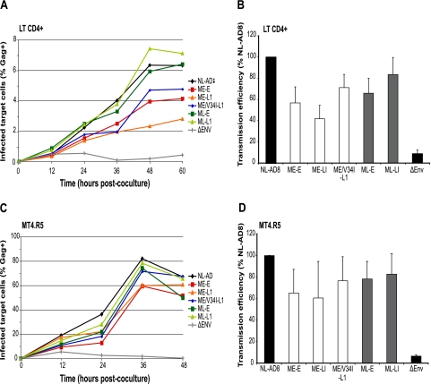
Lentiviruses, including human immunodeficiency virus type 1 (HIV-1), typically encode envelope glycoproteins (Env) with long cytoplasmic tails (CTs). The strong conservation of CT length in primary isolates of HIV-1 suggests that this factor plays a key role in viral replication and persistence in infected patients. However, we report here the emergence and dominance of a primary HIV-1 variant carrying a natural 20-amino-acid truncation of the CT in vivo. We demonstrated that this truncation was deleterious for viral replication in cell culture. We then identified a compensatory amino acid substitution in the matrix protein that reversed the negative effects of CT truncation. The loss or rescue of infectivity depended on the level of Env incorporation into virus particles. Interestingly, we found that a virus mutant with defective Env incorporation was able to spread by cell-to-cell transfer. The effects on viral infectivity of compensation between the CT and the matrix protein have been suggested by in vitro studies based on T-cell laboratory-adapted virus mutants, but we provide here the first demonstration of the natural occurrence of similar mechanisms in an infected patient. Our findings provide insight into the potential of HIV-1 to evolve in vivo and its ability to overcome major structural alterations.
Figures
Similar articles
-
HIV-1 Matrix Trimerization-Impaired Mutants Are Rescued by Matrix Substitutions That Enhance Envelope Glycoprotein Incorporation.J Virol. 2019 Dec 12;94(1):e01526-19. doi: 10.1128/JVI.01526-19. Print 2019 Dec 12. J Virol. 2019. PMID: 31619553 Free PMC article.
-
Rescue of human immunodeficiency virus type 1 matrix protein mutants by envelope glycoproteins with short cytoplasmic domains.J Virol. 1995 Jun;69(6):3824-30. doi: 10.1128/JVI.69.6.3824-3830.1995. J Virol. 1995. PMID: 7745730 Free PMC article.
-
Impact of natural polymorphism within the gp41 cytoplasmic tail of human immunodeficiency virus type 1 on the intracellular distribution of envelope glycoproteins and viral assembly.J Virol. 2007 Jan;81(1):125-40. doi: 10.1128/JVI.01659-06. Epub 2006 Oct 18. J Virol. 2007. PMID: 17050592 Free PMC article.
-
Domains of the human immunodeficiency virus type 1 matrix and gp41 cytoplasmic tail required for envelope incorporation into virions.J Virol. 1996 Jan;70(1):341-51. doi: 10.1128/JVI.70.1.341-351.1996. J Virol. 1996. PMID: 8523546 Free PMC article.
-
Interactions of the cytoplasmic domains of human and simian retroviral transmembrane proteins with components of the clathrin adaptor complexes modulate intracellular and cell surface expression of envelope glycoproteins.J Virol. 1999 Feb;73(2):1350-61. doi: 10.1128/JVI.73.2.1350-1361.1999. J Virol. 1999. PMID: 9882340 Free PMC article.
Cited by
-
Cell-Free versus Cell-to-Cell Infection by Human Immunodeficiency Virus Type 1 and Human T-Lymphotropic Virus Type 1: Exploring the Link among Viral Source, Viral Trafficking, and Viral Replication.J Virol. 2016 Aug 12;90(17):7607-17. doi: 10.1128/JVI.00407-16. Print 2016 Sep 1. J Virol. 2016. PMID: 27334587 Free PMC article. Review.
-
The Envelope Cytoplasmic Tail of HIV-1 Subtype C Contributes to Poor Replication Capacity through Low Viral Infectivity and Cell-to-Cell Transmission.PLoS One. 2016 Sep 6;11(9):e0161596. doi: 10.1371/journal.pone.0161596. eCollection 2016. PLoS One. 2016. PMID: 27598717 Free PMC article.
-
Multiple Gag domains contribute to selective recruitment of murine leukemia virus (MLV) Env to MLV virions.J Virol. 2013 Feb;87(3):1518-27. doi: 10.1128/JVI.02604-12. Epub 2012 Nov 14. J Virol. 2013. PMID: 23152533 Free PMC article.
-
Resistance of human immunodeficiency virus type 1 to a third-generation fusion inhibitor requires multiple mutations in gp41 and is accompanied by a dramatic loss of gp41 function.J Virol. 2011 Oct;85(20):10785-97. doi: 10.1128/JVI.05331-11. Epub 2011 Aug 10. J Virol. 2011. PMID: 21835789 Free PMC article.
-
The cytoplasmic tail of retroviral envelope glycoproteins.Prog Mol Biol Transl Sci. 2015;129:253-84. doi: 10.1016/bs.pmbts.2014.10.009. Epub 2014 Dec 1. Prog Mol Biol Transl Sci. 2015. PMID: 25595807 Free PMC article. Review.
References
-
- Altfeld, M., E. T. Kalife, Y. Qi, H. Streeck, M. Lichterfeld, M. N. Johnston, N. Burgett, M. E. Swartz, A. Yang, G. Alter, X. G. Yu, A. Meier, J. K. Rockstroh, T. M. Allen, H. Jessen, E. S. Rosenberg, M. Carrington, and B. D. Walker. 2006. HLA alleles associated with delayed progression to AIDS contribute strongly to the initial CD8+ T cell response against HIV-1. PLoS Med. 3:e403. - PMC - PubMed
-
- Amara, A., A. Vidy, G. Boulla, K. Mollier, J. Garcia-Perez, J. Alcami, C. Blanpain, M. Parmentier, J. L. Virelizier, P. Charneau, and F. Arenzana-Seisdedos. 2003. G protein-dependent CCR5 signaling is not required for efficient infection of primary T lymphocytes and macrophages by R5 human immunodeficiency virus type 1 isolates. J. Virol. 77:2550-2558. - PMC - PubMed
-
- Ataman-Onal, Y., C. Coiffier, A. Giraud, A. Babic-Erceg, F. Biron, and B. Verrier. 1999. Comparison of complete env gene sequences from individuals with symptomatic primary HIV type 1 infection. AIDS Res. Hum. Retrovir. 15:1035-1039. - PubMed
Publication types
MeSH terms
Substances
LinkOut - more resources
Full Text Sources
Research Materials
