. 2009 Sep;20(18):3996-4009.
doi: 10.1091/mbc.e09-05-0388. Epub 2009 Jul 22.

Role for RACK1 orthologue Cpc2 in the modulation of stress response in fission yeast

Affiliations

Role for RACK1 orthologue Cpc2 in the modulation of stress response in fission yeast

Andrés Núñez et al. Mol Biol Cell. 2009 Sep.

Abstract

The receptor of activated C kinase (RACK1) is a protein highly conserved among eukaryotes. In mammalian cells, RACK1 functions as an adaptor to favor protein kinase C (PKC)-mediated phosphorylation and subsequent activation of c-Jun NH(2)-terminal kinase mitogen-activated protein kinase. Cpc2, the RACK1 orthologue in the fission yeast Schizosaccharomyces pombe, is involved in the control of G2/M transition and interacts with Pck2, a PKC-type protein member of the cell integrity Pmk1 mitogen-activated protein kinase (MAPK) pathway. Both RACK1 and Cpc2 are structural components of the 40S ribosomal subunit, and recent data suggest that they might be involved in the control of translation. In this work, we present data supporting that Cpc2 negatively regulates the cell integrity transduction pathway by favoring translation of the tyrosine-phosphatases Pyp1 and Pyp2 that deactivate Pmk1. In addition, Cpc2 positively regulates the synthesis of the stress-responsive transcription factor Atf1 and the cytoplasmic catalase, a detoxificant enzyme induced by treatment with hydrogen peroxide. These results provide for the first time strong evidence that the RACK1-type Cpc2 protein controls from the ribosome the extent of the activation of MAPK cascades, the cellular defense against oxidative stress, and the progression of the cell cycle by regulating positively the translation of specific gene products involved in key biological processes.


Figures

Figure 1.
Figure 1.
Role of Cpc2 in the regulation of cell size, septation, and cell wall integrity is independent of the Pmk1 MAPK pathway. (A) Cell morphology and size at division (micrometers ± SD) in strains MI200 (control), AN100 (cpc2Δ), GB3 (pck2Δ), AN150 (pck2Δ cpc2Δ), TP319–13c (pmk1Δ), and AN160 (pmk1Δ cpc2Δ), growing in EMM2 medium after staining with calcofluor white. (B) Septation status of the cells in the above cultures (n ≥ 400). (C) The same strains were grown in YES medium (OD600 = 0.5) and assayed for β-glucanase sensitivity by treatment with 100 μg/ml Zymolyase 20-T at 30°C. Cell lysis was monitored by measuring decay in OD600 at different incubation periods, and the results shown are the mean value of three independent experiments. (D) Cell survival in the presence of Caspofungin. Samples containing 104, 103, 102 or 101 cells of wild type and single- and double-mutant strains grown in YES medium were spotted onto YES plates supplemented with 0, 0.7, or 1 μg/ml caspofungin and incubated for 3 d at 28°C before being photographed.
Figure 2.
Figure 2.
Cpc2 regulates chloride homeostasis in fission yeast alternatively to Pmk1. (A) Increased Pmk1 phosphorylation in cpc2Δ cells. Strains MI200 (pmk1-HA6H, control), AN100 (pmk1-HA6H, cpc2Δ), GB3 (pmk1-HA6H, pck2Δ), and AN150 (pmk1-HA6H, pck2Δ cpc2Δ) were grown in YES medium to mid-log phase, and Pmk1-HA6H was purified by affinity chromatography under native conditions. Activated and total Pmk1 was detected by immunoblotting with anti-phospho-p42/44 or anti-HA antibodies, respectively. (B) Chloride sensitivity assays for strains MI200 (control), GB3 (pck2Δ), TP319–13c (pmk1Δ), AN100 (cpc2Δ), AN150 (pck2Δ cpc2Δ), and AN160 (pmk1Δ cpc2Δ). After growth in YES medium, 104, 103, 102, or 101 cells were spotted onto YES plates supplemented with 0.2 M MgCl2 or 0.2 M MgCl2 plus 1 μg/ml FK506 and incubated for 3 d at 28°C before being photographed. (C) Strains MI200 (pmk1-HA6H, control), AN100 (pmk1-HA6H, cpc2Δ), MI212 (pmk1-HA6H, pmp1Δ), and AN170 (pmk1-HA6H, pmp1Δ cpc2Δ) were grown in YES medium to mid-log phase and total Pmk1-HA6H was purified and detected as described above. (D) Chloride sensitivity assays for strains MI200 (control), AN100 (cpc2Δ), PP42 (ppb1Δ), AN010 (ppb1Δ cpc2Δ), MI212 (pmp1Δ), and AN170 (pmp1Δ cpc2Δ). After growth in YES medium, cells were spotted onto YES plates supplemented with 75, 150, or 200 mM MgCl2 and incubated for 3 d at 28°C.
Figure 3.
Figure 3.
Cpc2 negatively regulates phosphorylation of Pmk1 and Sty1 MAPKs in growing and stressed cells. (A) Top, Strains MI200 (pmk1-HA6H, control), and AN100 (pmk1-HA6H, cpc2Δ), were grown in YES medium to mid-log phase and treated with 0.6 M KCl. At timed intervals Pmk1-HA6H was purified by affinity chromatography, and the activated and total Pmk1 was detected by immunoblotting with anti-phospho-p42/44 or anti-HA antibodies, respectively. Bottom, in the same experiment, Pmk1 phosphorylation was detected by immunoblotting with anti-phosphotyrosine antibody. (B) Strain AN100 (pmk1-HA6H, cpc2Δ) transformed with pREP-cpc2+ plasmid was grown for 18 h in the presence (+B1) or absence (−B1) of thiamine and treated with 0.6 M KCl. At different times, Pmk1-HA6H was purified and the activated, and total Pmk1 was detected as described in A. Cell morphology was analyzed by fluorescence microscopy. (C) Strains MI200 (pmk1-HA6H, control) and AN100 (pmk1-HA6H, cpc2Δ) were grown in YES medium to mid-log phase, treated with 1 mM H2O2, and the activated and total Pmk1 was detected as described in A. (D) Strains JM1521 (sty1-HA6H, control) and AN200 (sty1-HA6H, cpc2Δ) were grown in YES medium to mid-log phase and treated with 0.6 M KCl. At timed intervals, Sty1-HA6H was purified by affinity chromatography and the activated and total Sty1 was detected by immunoblotting with anti-phosphotyrosine antibody or anti-HA antibodies, respectively.
Figure 4.
Figure 4.
Cpc2 acts as a positive regulator of Pyp1 and Pyp2 tyrosine phosphatases protein levels. (A) Deletion of the cpc2+ gene in a wis1DD background rescues the defective Pmk1 activation in single wis1DD mutant. Strains MI200 (pmk1-HA6H, control), MI709 (pmk1-HA6H, wis1DD), AN600 (pmk1-HA6H, wis1DD cpc2Δ), and MI713 (pmk1-HA6H, wis1DD atf1Δ) were grown in YES medium to mid-log phase and treated with 0.6 M KCl. At the times indicated, aliquots were harvested and Pmk1-HA6H was purified by affinity chromatography. Activated Pmk1 was detected by immunoblotting with anti-phospho-p42/44 and total Pmk1 with anti-HA antibodies. (B) Protein levels of different MAPK phosphatases. The strains MI701 (pyp1–13myc, control) and AN700 (pyp1–13myc, cpc2Δ), MI702 (pyp2–13myc, control) and AN400 (pyp2–13myc, cpc2Δ), MI703 (ptc1–13myc, control) and AN500 (ptc1–13myc, cpc2Δ), AN030 (ptc3–13myc, control), and AN031 (ptc3–13myc, cpc2Δ), MI305 (pmp1-GFP, control) and AN020 (pmp1-GFP, cpc2Δ) were grown in YES medium to mid-log phase and treated with 0.6 M KCl. Total extracts were obtained from aliquots harvested at the times indicated, and fusions to 13-myc and GFP were detected by immunoblotting with either anti-c-myc or anti-GFP antibodies, respectively, whereas anti-Cdc2 antibody was used for loading control. (C) Defective synthesis of Pyp2 in cpc2Δ cells subjected to other stresses. Strains MI702 (pyp2–13myc, control) and AN400 (pyp2–13myc, cpc2Δ) were grown as described above and treated with 1 mM H2O2 (top) or incubated at 40°C (bottom). Aliquots were harvested at the times indicated and Pyp2–13myc was detected by immunoblotting with anti-c-myc antibody. Anti-Cdc2 antibody was used for loading control. (D) The basal Pmk1 phosphorylation in pyp1Δ cpc2Δ cells is similar to that of the single pyp1Δ mutant. Strains MI200 (pmk1-HA6H, control), AN100 (pmk1-HA6H, cpc2Δ), MI213 (pmk1-HA6H, pyp1Δ), and AN032 (pmk1-HA6H, pyp1Δ cpc2Δ), were grown as indicated previously and Pmk1-HA6H was purified by affinity chromatography under native conditions. Activated and total Pmk1 was detected by immunoblotting with anti-phospho-p42/44 or anti-HA antibodies, respectively.
Figure 5.
Figure 5.
The protein level of Atf1 is positively regulated by Cpc2. (A) Strains JM1821 (atf1-HA6H, control) and AN300 (atf1-HA6H, cpc2Δ) were grown in YES medium to mid-log phase and treated with either 0.6 M KCl (top) or 1 mM H2O2 (bottom). Aliquots were harvested at different times, and Atf1 was purified from cell extracts by affinity chromatography, and detected by immunoblotting with anti-HA antibody. Anti-Cdc2 antibody was used for loading control. (B) Strains JM1821 and AN300 were grown at 28°C in YES medium to mid-exponential phase and treated with 0.6 M KCl for the times indicated. Total RNA was extracted, denatured, transferred to nylon membranes, and hybridized with 32P-labeled probes for atf1+, pyp2+, pyp1+, and leu1+ (loading control). (C) These same strains were grown in YES medium (OD600 = 0.5) and treated with 100 μg/ml cycloheximide for the times indicated. Purified Atf1 was detected by immunoblotting with anti-HA antibody, whereas anti-α-tubulin antibody was used as loading control. (D) EIF2α is not involved in the control of Atf1 translation by Cpc2. Strains JM1821 (atf1-HA6H, control), AN300 (atf1-HA6H, cpc2Δ), AN040 (atf1-HA6H, EIF2α-S52A), and AN041 (atf1-HA6H, EIF2α-S52A cpc2Δ) were grown in YES medium and treated with either 0.6 M KCl (top) or 1 mM H2O2 (bottom). Atf1 from each sample was purified by affinity chromatography and detected by immunoblotting with anti-HA antibody. Anti-Cdc2 antibody was used for loading control.
Figure 6.
Figure 6.
Cellular defense against hydrogen peroxide is defective in the absence of Cpc2. (A) Cell viability assays in solid medium were performed for strains MI200 (control), AN100 (cpc2Δ), TK107 (sty1Δ), and MI103 (atf1Δ). After growth in YES medium, 104, 103, 102, or 101 cells were spotted onto YES plates supplemented with either 0.6 M KCl, 1.2 M sorbitol, or 1.5–2 mM H2O2 and incubated for 4 d at 28°C before being photographed. (B) Cell viability assays in liquid medium for the above strains after growth in YES liquid medium (OD600 = 0.5) and treatment with 5 mM H2O2 for 30 min before seeding in YES plates. Survival was estimated as percentage of colony forming units relative to untreated cultures. The experiment was repeated three times with similar results. (C) Nuclear accumulation of GFP-Pap1 is delayed in cpc2Δ cells. Strains EHH14 (GFP-Pap1; control) and AN150 (GFP-Pap1, cpc2Δ), expressing a GFP-Pap1 fusion under the control of the nmt1-promoter, were grown in EMM2 medium to an OD600 = 0.5 and treated with 1 mM H2O2. Samples were taken at different times, and the percentage of cells with nuclear accumulation of GFP-Pap1 was determined by fluorescence microscopy. Results shown correspond to one representative experiment.
Figure 7.
Figure 7.
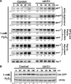
Cpc2 modulates S. pombe response to oxidative stress at both transcriptional and translational levels. (A) Induced expression of Pap1-dependent genes in response to hydrogen peroxide is delayed in cpc2Δ cells. Strains MI200 (control) and AN100 (cpc2Δ) were grown in YES medium to early log phase and treated with 1 mM H2O2 for the times indicated. Total RNA was extracted from each sample and 20 μg resolved in 1.5% agarose formaldehyde gels. The denatured RNAs were transferred to nylon membranes and hybridized with 32P-labeled probes for atf1+, pyp2+, gpx1+, srx1+, ctt1+, tpx1+, trr1+, and leu1+ (loading control). (B) Increased synthesis of cytoplasmic catalase in response to hydrogen peroxide is blocked in cpc2Δ cells. Strains AN060 (control; ctt1-GFP) and AN061 (cpc2Δ, ctt1-GFP) were grown in YES medium and treated with 1 mM H2O2 for the times indicated. Total extracts were obtained and Ctt1-GFP fusion was detected by immunoblotting with anti-GFP antibodies. Anti-Cdc2 antibody was used for loading control.
Figure 8.
Figure 8.
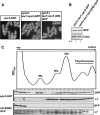
Construction of a S. pombe strain expressing a Cpc2 version unable to bind the ribosome in vivo. (A) Subcellular localization of Cpc2 variants. Mid-log phase cells of strains AN070 (cpc2-GFP, control), AN071 (leu1:cpc2-GFP, cpc2Δ), and AN072 (leu1:cpc2(DE)-GFP, cpc2Δ) growing in YES medium were visualized by fluorescence microscopy. (B) The Cpc2-GFP and Cpc2(DE)-GFP fusions are expressed at a similar level. GFP fusions were detected in total cell extracts from strains AN071 (leu1:cpc2-GFP, cpc2Δ) and AN072 (leu1:cpc2(DE)-GFP, cpc2Δ) by immunoblotting with anti-GFP antibodies. Anti-Cdc2 antibody was used for loading control. (C) Polysome Western blot analysis for in vivo ribosome binding. Strains AN071 and AN072 were grown in YES medium to early log phase, treated with cycloheximide, and the whole cell extracts processed for polysome analysis by velocity sedimentation on a 7–47% (wt/vol) sucrose gradient. After fractionation, the absorbance profile for each fraction was measured at 254 nm. A typical result is shown, with fractions corresponding to soluble (top), 40S, 60S, 80S, and polyribosomes. Aliquots from each fraction were resolved by SDS-PAGE and the elution profile for Cpc2-GFP or Cpc2 (DE)-GFP was determined by Western blot analysis with anti-GFP antibody. Anti-L7 antibody was used as an internal ribosomal control.
Figure 9.
Figure 9.
Ribosome binding of Cpc2 is essential to positively regulate the translation efficiency of Pyp1, Pyp2, and Atf1 in fission yeast. (A) Increased Pmk1 phosphorylation in cells expressing a Cpc2(DE) version. Strains AN071 (pmk1-HA6H leu1:cpc2-GFP, cpc2Δ) and AN072 (pmk1-HA6H leu1:cpc2(DE)-GFP, cpc2Δ) were grown to mid-log phase in YES medium and treated with 0.6 M KCl for the indicated times. Pmk1-HA6H was purified by affinity chromatography under native conditions and the activated and total Pmk1 detected by immunoblotting with anti-phospho-p42/44 or anti-HA antibodies, respectively. (B) Decreased Pyp1 and Pyp2 protein levels in the Cpc2(DE) mutant. Strains AN091 (leu1:cpc2-GFP cpc2Δ pyp1–13myc) and AN092 (leu1:cpc2(DE)-GFP cpc2Δ pyp1–13myc) (upper panel), or AN102 (leu1:cpc2-GFP cpc2Δ pyp2–13myc) and AN103 (leu1:cpc2(DE)-GFP cpc2Δ pyp2–13myc) (lower panel) were grown in YES medium to mid-log-phase and treated with 0.6 M KCl. Aliquots were harvested at the times indicated and total extracts obtained. Pyp1–13myc or Pyp2–13myc fusions were detected by immunoblotting with anti-c-myc antibody. Anti-Cdc2 antibody was used for loading control. (C) Decreased Atf1 protein levels in the Cpc2(DE) mutant. Strains AN081 (atf1-HA6H leu1:cpc2-GFP cpc2Δ) and AN082 (atf1-HA6H leu1:cpc2(DE)-GFP cpc2Δ) were grown as above and treated with either 0.6 M KCl (top) or 1 mM H2O2 (bottom). Atf1 was purified by affinity chromatography from cell extracts of aliquots harvested at different time and detected by immunoblotting with anti-HA antibody. Anti-Cdc2 antibody was used for loading control. (D) cpc2Δ cells expressing the Cpc2(DE) version do not recover normal cell size at division. Cell morphology and size in strains AN100 (cpc2Δ), AN071 (leu1:cpc2-GFP, cpc2Δ), and AN072 (leu1:cpc2(DE)-GFP, cpc2Δ) growing in EMM2 medium and stained with calcofluor white are shown.
Figure 10.
Figure 10.
Proposed functional role for RACK orthologue Cpc2 in fission yeast. The Cpc2 protein regulates important biological processes at the ribosome level by favoring the translation of genes whose products may inactivate MAPK cascades (Pyp1 and Pyp2 MAPK phosphatases), enhance cellular defense against oxidative stress (transcription factor Atf1, cytoplasmic catalase Ctt1), and control G2/M transition of the cell cycle. The question mark highlights that the additional role of one or several proteins is not discarded.

Similar articles

Cited by

References

    1. Alfa C., Fantes P., Hyams J., Mcleod M., Warbrick E. Experiments with Fission Yeast. A Laboratory Course Manual. Cold Spring Harbor, NY: Cold Spring Harbor Laboratory Press; 1993.
    1. Bahler J., Wu J. Q., Longtine M. S., Shah N. G., McKenzie A., 3rd, Steever A. B., Wach A., Philippsen P., Pringle J. R. Heterologous modules for efficient and versatile PCR-based gene targeting in Schizosaccharomyces pombe. Yeast. 1998;14:943–951. - PubMed
    1. Barba G., Soto T., Madrid M., Núñez A., Vicente J., Gacto M., Cansado J. Activation of the cell integrity pathway is channelled through diverse signalling elements in fission yeast. Cell. Signal. 2008;20:748–757. - PubMed
    1. Baum S., Bittins M., Frey S., Seedorf M. Asc1p, a WD40-domain containing adaptor protein, is required for the interaction of the RNA-binding protein Scp160p with polysomes. Biochem. J. 2004;380:823–830. - PMC - PubMed
    1. Benko Z., Sipiczki M., Carr A. M. Cloning of caf1+, caf2+ and caf4+ from Schizosaccharomyces pombe: their involvement in multidrug resistance, UV and pH sensitivity. Mol. Gen. Genet. 1998;260:434–443. - PubMed

Publication types

MeSH terms

LinkOut - more resources