. 2009 Sep;37(17):5678-89.
doi: 10.1093/nar/gkp593. Epub 2009 Jul 22.

Inhibition of ATR protein kinase activity by schisandrin B in DNA damage response

Affiliations

Inhibition of ATR protein kinase activity by schisandrin B in DNA damage response

Hiroshi Nishida et al. Nucleic Acids Res. 2009 Sep.

Abstract

ATM and ATR protein kinases play a crucial role in cellular DNA damage responses. The inhibition of ATM and ATR can lead to the abolition of the function of cell cycle checkpoints. In this regard, it is expected that checkpoint inhibitors can serve as sensitizing agents for anti-cancer chemo/radiotherapy. Although several ATM inhibitors have been reported, there are no ATR-specific inhibitors currently available. Here, we report the inhibitory effect of schisandrin B (SchB), an active ingredient of Fructus schisandrae, on ATR activity in DNA damage response. SchB treatment significantly decreased the viability of A549 adenocarcinoma cells after UV exposure. Importantly, SchB treatment inhibited both the phosphorylation levels of ATM and ATR substrates, as well as the activity of the G2/M checkpoint in UV-exposed cells. The protein kinase activity of immunoaffinity-purified ATR was dose-dependently decreased by SchB in vitro (IC(50): 7.25 muM), but the inhibitory effect was not observed in ATM, Chk1, PI3K, DNA-PK, and mTOR. The extent of UV-induced phosphorylation of p53 and Chk1 was markedly reduced by SchB in ATM-deficient but not siATR-treated cells. Taken together, our demonstration of the ability of SchB to inhibit ATR protein kinase activity following DNA damage in cells has clinical implications in anti-cancer therapy.


Figures

Figure 1.
Figure 1.
Chemical structure of SchB.
Figure 2.
Figure 2.
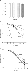
SchB treatment causes a decrease in cell viability after UV irradiation. Relative cell viability was measured by clonogenic assay. A549 cells were seeded as 500 cells/60-mm dish, and then incubated for 24 h before the experiment. The cells were subjected to 1 h pre-incubation and 14 days post-incubation (A) with SchB (0, 1, 10 and 30 μM) after (B) UV irradiation (0, 20 and 50 J/m2) or (C) IR irradiation (0, 2 and 6 Gy). Values are expressed as mean ± SD (n = 3). *P < 0.05 versus SchB untreated control.
Figure 3.
Figure 3.
SchB treatment abolishes cell cycle checkpoints induced by UV irradiation. (A) Effect of SchB on the G2/M checkpoint after UV-induced DNA damage using G2/M synchronized cells. Cell-cycle distributions were analyzed by FACS after 1 h of UV exposure, followed by data analysis using ModFit software. The histogram in gray represents cells synchronized at the G2/M phase by nocodazole. A549 cells were treated with 30 μM SchB or 10 mM caffeine 1 h before UV irradiation (0, 20 and 50 J/m2). The arrow shows the cells distributed in the G1 phase. (B) Data were expressed as the percentage of G1 cells in relation to the total number of cells. (C) The percentage of mitotic cells was estimated by phosphorylation of histone H3 at Ser10. (D) The mitotic percentage of asynchronous cells was estimated by phosphorylation of histone H3 in 10 J/m2 of UV irradiation followed by 1h incubation. (E) The percentage of DNA synthesizing cells was visualized by BrdU intake of cells. (F) Data obtained in Figure 3E were analyzed using XL2 software to determine the percentage of mitotic cells. Values are expressed as mean ± SD (n = 3). *P < 0.05 versus SchB untreated control.
Figure 4.
Figure 4.
Inhibitory effects of SchB treatment on checkpoint proteins. (A) A549 cells were pre-incubated with or without 30 μM SchB for 1 h prior to induction of DNA damage. After pre-incubation, cells were subjected to 10 Gy of IR or 20 J/m2 of UV irradiation, and cultured at 37°C for 1 h and 3 h, respectively. (B) Dose-dependent effects of SchB on the phosphorylation level of p53 and Chk1 were observed by adding increasing concentrations of SchB before and after induction of DNA damage by UV irradiation (20 J/m2). A549 cells were incubated with a proteasome inhibitor (50 μM LLnL) throughout the experiments in A, B and C to avoid proteasome-dependent protein degradation. Equal loading of extracted proteins was confirmed by determining immuno-stained tubulin. (C) A549 cells were pre-incubated with or without 30 μM SchB or 10 mM caffeine for 1 h prior to induction of DNA damage. After pre-incubation, cells were subjected to UV irradiation (20 J/m2), and cultured at 37°C for 3 h. (D) Flag-tagged ATR was expressed in HEK293T cells following transient transfection with a flag-tagged ATR, and the expressed proteins were immunoprecipitated. The association of ATR with ATRIP was analyzed by immunoblot analysis.
Figure 5.
Figure 5.
SchB treatment inhibits ATR kinase activity. Kinase activities of ATR (A), ATM (B), Chk1 (C), PI3K (D), DNA-PK (E) and mTOR (F) were measured in vitro as phosphorylation activity in the presence of SchB. A flag-tagged ATR-wt plasmid was transfected into HEK293T cells. ATR protein kinases were purified by immunoprecipitation using anti-Flag M2 antibody and Protein-G agarose. The endogeneous ATM protein was purified from A549 cell lysates with anti-ATM antibody. Kinase activity was monitored for 20 min at 30°C. Chk1 (Upstate), PI3K (Echelon Biosciences), DNA-PK (Promega) and mTOR (Calbiochem) activities were measured using respective assay kits according to the manufacturers instructions. Values are presented as mean ± SD (n = 3).
Figure 6.
Figure 6.
SchB prevents ATR-dependent signaling pathway after DNA damage. (A) AT2KY fibroblasts were pre-incubated with or without 30 μM SchB for 1 h, and then subjected to UV irradiation (20 J/m2). The cells were harvested 4 h later and phosphorylation levels of p53, Chk1, SMC1 and BRCA1 were measured to assess checkpoint function. (B) A549 cells were transfected with siRNA for control (siGFP), ATM or ATR using the oligofectamine method, and the cell extracts were immunoblotted with anti-phosphorylated p53, phosphorylated Chk1, total-ATM, total-ATR or anti-tubulin. After 72 h incubation for the knockdown of ATM or ATR, cells were subjected to UV irradiation (20 J/m2) followed by 4 h incubation with or without SchB (30 μM) pre/post-incubation. The equal loading of an extracted protein was confirmed using anti-monoclonal tubulin antibody. (C) The cell viability was measured in ATR-deficient Seckel syndrome fibroblasts with or without SchB (0, 1, 10 and 30 μM) after UV irradiation (0, 25, 50 and 75 J/m2). Values are expressed as mean ± SD (n = 3).
Figure 7.
Figure 7.
Phosphorylation of ERK at Thr202 and Tyr204. A549 cells were pre-treated with 50 μM PD098059 or 30 μM SchB after serum starvation for 16 h. The phosphorylation of ERK was activated by adding 100 ng/ml PMA followed by a 5 min incubation. The cells were lysed and applied to immunoblot analysis. Equal loading of an extracted protein was confirmed using anti-monoclonal tubulin antibody.

References

    1. Shiloh Y. ATM and ATR: networking cellular responses to DNA damage. Curr. Opin. Genet. Dev. 2001;11:71–77. - PubMed
    1. Abraham RT. Cell cycle checkpoint signaling through the ATM and ATR kinases. Genes Dev. 2001;15:2177–2196. - PubMed
    1. Collis SJ, Swartz MJ, Nelson WG, DeWeese TL. Enhanced radiation and chemotherapy-mediated cell killing of human cancer cells by small inhibitory RNA silencing of DNA repair factors. Cancer Res. 2003;63:1550–1554. - PubMed
    1. Brown EJ, Baltimore D. Essential and dispensable roles of ATR in cell cycle arrest and genome maintenance. Genes Dev. 2003;17:615–628. - PMC - PubMed
    1. Matsuoka S, Ballif BA, Smogorzewska A, McDonald ER, III, Hurov KE, Luo J, Bakalarski CE, Zhao Z, Solimini N, Lerenthal Y, et al. ATM and ATR substrate analysis reveals extensive protein networks responsive to DNA damage. Science. 2007;316:1160–1166. - PubMed

Publication types

MeSH terms