. 2009 Aug 1;122(Pt 15):2686-98.
doi: 10.1242/jcs.035287.

Role of cleavage by separase of the Rec8 kleisin subunit of cohesin during mammalian meiosis I

Affiliations

Role of cleavage by separase of the Rec8 kleisin subunit of cohesin during mammalian meiosis I

Nobuaki R Kudo et al. J Cell Sci. .

Abstract

Proteolytic activity of separase is required for chiasma resolution during meiosis I in mouse oocytes. Rec8, the meiosis-specific alpha-kleisin subunit of cohesin, is a key target of separase in yeast. Is the equivalent protein also a target in mammals? We show here that separase cleaves mouse Rec8 at three positions in vitro but only when the latter is hyper-phosphorylated. Expression of a Rec8 variant (Rec8-N) that cannot be cleaved in vitro at these sites causes sterility in male mice. Their seminiferous tubules lack a normal complement of 2 C secondary spermatocytes and 1 C spermatids and contain instead a high proportion of cells with enlarged nuclei. Chromosome spreads reveal that Rec8-N expression has no effect in primary spermatocytes but produces secondary spermatocytes and spermatids with a 4 C DNA content, suggesting that the first and possibly also the second meiotic division is abolished. Expression of Rec8-N in oocytes causes chromosome segregation to be asynchronous and delays its completion by 2-3 hours during anaphase I, probably due to inefficient proteolysis of Rec8-N by separase. Despite this effect, chromosome segregation must be quite accurate as Rec8-N does not greatly reduce female fertility. Our data is consistent with the notion that Rec8 cleavage is important and probably crucial for the resolution of chiasmata in males and females.


Figures
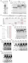
Fig. 1.
Fig. 1.
Identification of separase cleavage sites within Rec8 in vitro. [35S]Methionine-labeled mouse wild-type and mutant Rec8 and human SCC1-Myc were prepared in an in vitro transcription-translation system (IVT) and subjected to an in vitro separase cleavage assay using recombinant active separase (lanes marked A) or its inactive mutant (lanes marked i) in the presence or absence of recombinant polo-like kinase 1 (Plk1) as indicated. Autoradiographs of SDS-PAGE gels on which incubated samples were run are shown. Fragments resulting from cleavage at the primary, secondary and tertiary sites are indicated by asterisks and brackets in red, blue and green, respectively. (A) Cleavage assay of mouse Rec8 and C-terminally 9× Myc-tagged human SCC1. Arrows indicate full-length proteins. Cleavage fragments (asterisks) are the bands uniquely seen in A. (B) Cleavage assay in the presence of Plk1. Duplicated samples were run side by side. The amount (μg) of purified Plk1 added to each reaction is indicated. Dashed line (right) shows positions of phosphorylated Rec8. (C) Identities of mouse Rec8 mutants. (D) Alignment of experimentally proven separase target sites (Uhlmann et al., 1999; Buonomo et al., 2000; Tomonaga et al., 2000; Hauf et al., 2001; Sullivan et al., 2001; Waizenegger et al., 2002; Zou et al., 2002; Kitajima et al., 2003). The arrowhead indicates positions of cleavage. Mm, Mus musculus; Hs, Homo sapiens; Sc, Saccharomyces cerevisiae; Sp, Schizosaccharomyces pombe. (E) Cleavage assay of wild-type (WT), M1, M2, M3 and C-terminally 9× Myc-tagged wild-type Rec8 (Myc). (F) Cleavage assay of M2, M21 and M22. (G) Cleavage assay of WT, M2, M2/4, M2/5 and M2/4/5. (H) Cleavage assay of M2, M2/5, M2/5/6 and M2/5/7.
Fig. 2.
Fig. 2.
Male transgenic mice expressing Rec8-N-Myc fail to produce haploid cells. (A) A schematic representation of the mouse Rec8 genomic locus showing the mutagenized loci for generating BAC-mediated transgene constructs. A DNA fragment encoding 9× Myc epitopes (9× Myc) was inserted at the termination codon (asterisk) to make the Rec8-Myc expression construct (for TG Rec8-Myc mice). This construct was further modified to obtain the Rec8-N-Myc expression construct (for TG Rec8-N-Myc mice) by replacing the following residues: 262nd Arg to Asp (R262D), 434th Arg to Asp (R434D) and 454th Arg to Glu (R454E). For a conditional construct (for TG Stop/Rec8-N-Myc mice), a loxP-flanked stop cassette was inserted at the intron 1 (4× polyA). Boxes represent exons. ATG, initiation codon; S, Stu I site; The P represents the position of cleavage sites in Fig. 1C. (B) Southern blot analysis of genomic DNA from transgenic mice and littermates without transgene (No TG) digested by Stu I. The BACs encoding Rec8 or Rec8-Myc were examined as controls (BAC). The 1089 bp Stu I fragment was used as a probe. Expected sizes of fragments encoding the C-termini of Rec8 and Rec8-Myc are 1089 (green arrowhead) and 1478 bp (red arrowhead), respectively. (C) Western blot analysis testing the expression of Rec8-Myc and Rec8-N-Myc in total testicular cell extracts. Genotypes of endogenous Rec8 are indicated. One blot was used for three examinations by anti-Rec8, anti-Myc and anti-tubulin (loading control). Green arrowhead, endogenous Rec8; red arrowhead, Rec8-Myc or Rec8-N-Myc. (D) FACS analysis of testicular cells for the DNA content. Histogram peaks indicated as 1C, 2C and 4C correspond to cells having one, two or four copies of the genome, respectively.
Fig. 3.
Fig. 3.
Spermatogenic failure of TG Rec8-N-Myc males. Hematoxylin and eosin staining of seminiferous tubule sections from transgenic mice (TG) and a non-transgene littermate (No TG). Representative sections of stage V (A) and XII (B) are shown. Representative nuclei for each cell type present in the sections are shown at higher magnification. Scale bars: 20 and 5 μm for low and high magnification images, respectively.
Fig. 4.
Fig. 4.
Morphological change of Sycp3 after meiosis I is unaffected in TG Rec8-N-Myc spermatocytes. Immunofluorescence images of stage XII tubule sections stained by DAPI (for DNA) and anti-Sycp3 antibody. Arrowheads and arrows indicate typical patterns of Sycp3 signals in metaphase I and post-metaphase I, respectively. Stages are: Z, zygotene; MI, metaphase I; Int, interkinesis; 12, step 12 elongating spermatid. Scale bars: 10 μm.
Fig. 5.
Fig. 5.
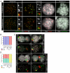
Normal chromosome development until metaphase I in TG Rec8-N-Myc spermatocytes. Immunofluorescence images of chromosome spreads stained with anti-Myc and anti-Sycp3 antibodies. (A) Pachytene nuclei and magnified sex chromosomes synapsed at the PAR (arrow) are shown. (B) Metaphase I chromosomes and magnified images of representative bivalents are shown.
Fig. 6.
Fig. 6.
No nuclear division in spermatocytes expressing Rec8-N-Myc. (A) Immunofluorescence images of chromosome spreads stained with a CREST serum (for centromeres), anti-Sycp3 antibody and DAPI (for DNA). Small panels show the magnified images of boxed areas. (B) Frequencies of nuclei classified by number of centromeres: approximately 40 CREST foci (40× Cen) or approximately 80 CREST foci (80× Cen). Numbers of nuclei examined are indicated (n). (C) Chromosome paintings detecting X (red) and Y (green) chromosomes merged on DAPI images or on anti-Sycp3 images in grayscale. Stages are: Pa, pachytene; Dip, diplotene; Int, interkinesis; RS, round spermatid. (D) Frequencies of nuclei classified by distribution of sex chromosome painting signals: one patch of X chromosome alone (X), one patch of Y chromosome alone (Y) or one or two patches of both (X and Y). Numbers of nuclei examined are indicated (n).
Fig. 7.
Fig. 7.
Double number of dyad chromosomes in metaphase II TG Rec8-N-Myc spermatocytes. (A) Condensed chromosomes found in Giemsa-stained spread preparations: 40 chromosomes with arm closed (Mitotic Metaphase); approximately 20 bivalents; approximately 20 dyads; approximately 40 dyads. (B) Frequencies of different classes of condensed chromosomes shown in A. Numbers of nuclei examined are indicated (n).
Fig. 8.
Fig. 8.
Persistence of Rec8-N-Myc on chromatin after meiosis I and II. Immunofluorescence images of chromosome spreads stained with anti-Myc and anti-Sycp3 antibodies. (A) Interkinesis nuclei and representative areas in higher magnification that contain centromeric Rec8 foci are shown. (B) Spermatid nuclei and representative chromocenters in higher magnification that possess Sycp3 signals are shown.
Fig. 9.
Fig. 9.
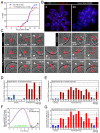
Rec8-N delays but does not block chiasma resolution at meiosis I in oocytes. (A) Kinetics of the first polar body extrusion (PBE) of oocytes cultured in vitro. The numbers of oocytes examined are indicated (n). (B) Localization of Rec8-N-Myc on chromosomes in TG Rec8-N-Myc oocytes. Chromosome spreads prepared from oocytes in culture for the indicated time after germinal vesicle breakdown (GVBD) were stained with anti-Myc antibody (red), CREST antiserum (green) and DAPI (blue). (C) Live confocal microscopy of oocytes expressing H2B-mCherry. DIC images (gray) were merged with images of the mCherry channel (red). Frames at indicated time (hours after beginning of filming) were selected from the original time series (supplementary material Movie 1). PBs are indicated by arrowheads or circles (when extruded vertically). (D) Intervals from PBE to the completion of chromosome segregation analyzed by ∼16-minute filming intervals (experiment A, supplementary material Movie 1). Error bar, s,d. (E) As in (D), analyzed by 5-7 minute intervals (experiment B, movies not shown). (F) Representative fluorescence quantifications of securin-EGFP signal in individual oocytes. The maximum value of the signal in each oocyte was set to 100 and relative intensity was plotted. Flags indicate time of the completion of chromosome segregation. Green line indicates the 5% level of the EGFP signal. (G) Intervals from 5% of the EGFP signal to the completion of chromosome segregation analyzed by 5-7 minute filming intervals (experiment B, movies not shown). Error bar, s.d.

Similar articles

Cited by

References

    1. Alexandru, G., Uhlmann, F., Mechtler, K., Poupart, M. A. and Nasmyth, K. (2001). Phosphorylation of the cohesin subunit Scc1 by Polo/Cdc5 kinase regulates sister chromatid separation in yeast. Cell 105, 459-472. - PubMed
    1. Bannister, L. A., Reinholdt, L. G., Munroe, R. J. and Schimenti, J. C. (2004). Positional cloning and characterization of mouse mei8, a disrupted allelle of the meiotic cohesin Rec8. Genesis 40, 184-194. - PubMed
    1. Brar, G. A., Kiburz, B. M., Zhang, Y., Kim, J. E., White, F. and Amon, A. (2006). Rec8 phosphorylation and recombination promote the step-wise loss of cohesins in meiosis. Nature 441, 532-536. - PubMed
    1. Buonomo, S. B., Clyne, R. K., Fuchs, J., Loidl, J., Uhlmann, F. and Nasmyth, K. (2000). Disjunction of homologous chromosomes in meiosis I depends on proteolytic cleavage of the meiotic cohesin Rec8 by separin. Cell 103, 387-398. - PubMed
    1. Clyne, R. K., Katis, V. L., Jessop, L., Benjamin, K. R., Herskowitz, I., Lichten, M. and Nasmyth, K. (2003). Polo-like kinase Cdc5 promotes chiasmata formation and cosegregation of sister centromeres at meiosis I. Nat. Cell Biol. 5, 480-485. - PubMed

Publication types

MeSH terms