. 2009 Jul 22;29(29):9137-47.
doi: 10.1523/JNEUROSCI.5478-08.2009.

Protocadherin-alpha family is required for serotonergic projections to appropriately innervate target brain areas

Affiliations

Protocadherin-alpha family is required for serotonergic projections to appropriately innervate target brain areas

Shota Katori et al. J Neurosci. .

Abstract

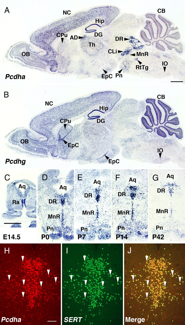
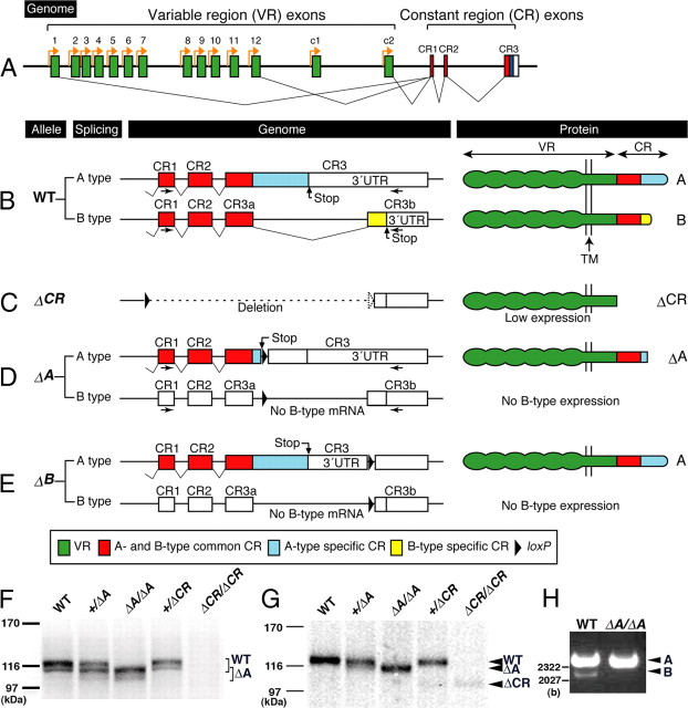
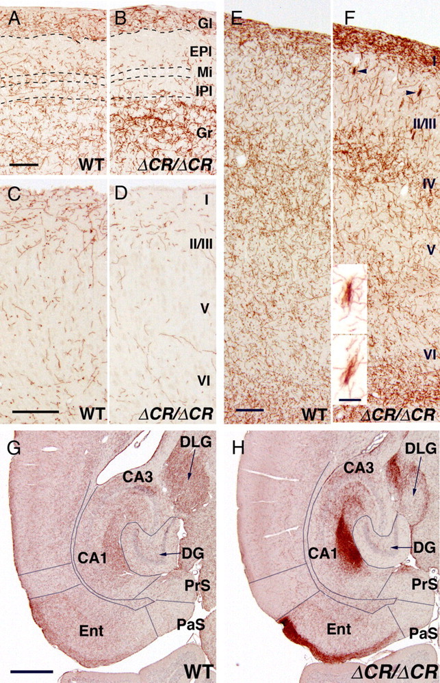
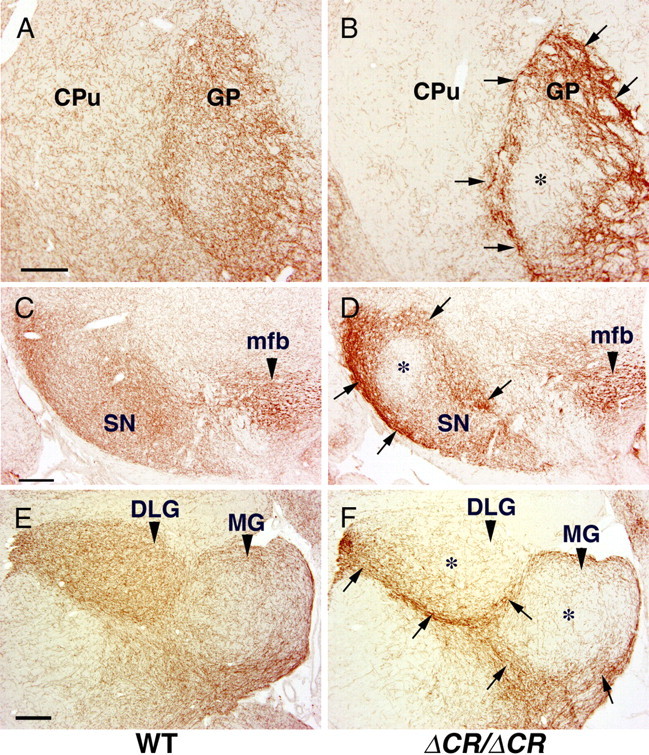
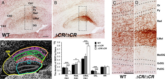
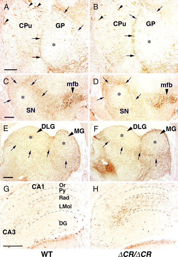
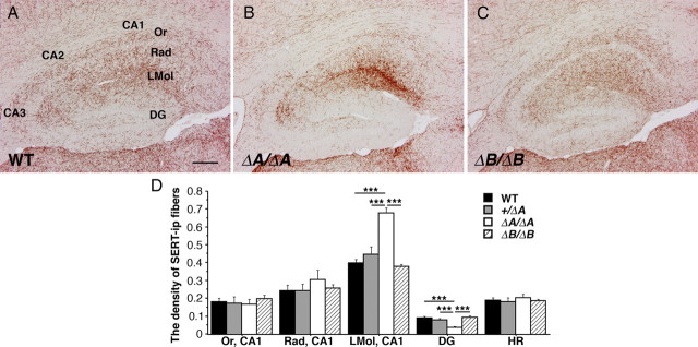
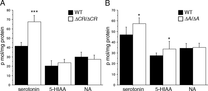
Serotonergic axons from the raphe nuclei in the brainstem project to every region of the brain, where they make connections through their extensive terminal arborizations. This serotonergic innervation contributes to various normal behaviors and psychiatric disorders. The protocadherin-alpha (Pcdha) family of clustered protocadherins consists of 14 cadherin-related molecules generated from a single gene cluster. We found that the Pcdhas were strongly expressed in the serotonergic neurons. To elucidate their roles, we examined serotonergic fibers in a mouse mutant (Pcdha(Delta CR/Delta CR)) lacking the Pcdha cytoplasmic region-encoding exons, which are common to the gene cluster. In the first week after birth, the distribution pattern of serotonergic fibers in Pcdha(Delta CR/Delta CR) mice was similar to wild-type, but by 3 weeks of age, when the serotonergic axonal termini complete their arborizations, the distribution of the projections was abnormal. In some target regions, notably the globus pallidus and substantia nigra, the normally even distribution of serotonin axonal terminals was, in the mutants, dense at the periphery of each region, but sparse in the center. In the stratum lacunosum-molecular of the hippocampus, the mutants showed denser serotonergic innervation than in wild-type, and in the dentate gyrus of the hippocampus and the caudate-putamen, the innervation was sparser. Together, the abnormalities suggested that Pcdha proteins are important in the late-stage maturation of serotonergic projections. Further examination of alternatively spliced exons encoding the cytoplasmic tail showed that the A-type (but not the B-type) cytoplasmic tail was essential for the normal development of serotonergic projections.


Figures

Figure 1.
Figure 1.
Serotonergic neurons strongly express Pcdhas in the mouse brain. A, B, The expression patterns of Pcdhas (A) and Pcdhgs (B) in adjacent sagittal sections of P14 mice. Pcdhas and Pcdhgs were strongly expressed in various regions, including the olfactory bulb (OB), neocortex (NC), hippocampus (Hip), dentate gyrus (DG), thalamus (Th), and cerebellum (CB). Pcdhas were very strongly expressed in the rostral raphe nuclei (DR, dorsal raphe nuclei; MnR, median raphe nucleus; CLi, caudal linear nucleus raphe). Pcdhgs were expressed in the caudate-putamen (CPu), ependymal cells (EpC), and inferior olive (IO). AD, Anterodorsal thalamic nucleus; Pn, pontine nuclei; RtTg, reticulotegmental nucleus of the pons. C–G, Coronal sections showing expression of Pcdhas in the rostral raphe nuclei at embryonic day (E) 14.5 (C), P0 (D), P7 (E), P14 (F), and P42 (G). The expression levels of Pcdhas were highest between E14.5 and P14. Aq, Aqueduct; Ra, raphe nuclei. H–J, Double fluorescent in situ hybridization with Pcdha-CR (H, arrowheads) and SERT probes (I, arrowheads) in the DR at P21 showed that SERT-positive cells were consistent with Pcdha-CR-positive cells (J, arrowheads). Scale bars: (A–G), 1 mm; (H–J), 200 μm.
Figure 2.
Figure 2.
Summary of Pcdha mutant alleles and their products. A, Pcdha genes consist of variable region (VR) exons (1–12, c1 and c2; green) and CR exons (CR1–CR3). The individual VR exons are transcribed from their own promoter (orange arrows). A Pcdha transcript is produced from one of the variable region exons and a set of CR exons by splicing. B, In the wild-type (WT) allele, alternative splicing of the CR exons of Pcdha genes produces A- and B-type variants (B, left). The CR exons consist of an A- and B-common coding region (red), an A-specific coding region (blue), a B-specific coding region (yellow), and a 3′UTR (white). Pcdha proteins are single-pass membrane proteins. The extracellular, transmembrane (TM) and cytoplasmic domains are encoded in the VR exons, and their cytoplasmic domain is encoded in the CR exons (B, right). C, In the ΔCR allele, the coding region of the CR exons was deleted (C, left). From the ΔCR allele, CR-deleted Pcdha proteins (ΔCR) were faintly expressed (C, right; F, G). D, In the ΔA allele, a termination codon and a loxP site were inserted into the A-specific coding region (D, left). From the ΔA allele, truncated proteins (ΔA) were expressed (D, right). E, In the ΔB allele, a loxP site was inserted into the B-specific coding region (E, left). From the ΔB allele, A-type proteins were normally expressed, but B-type proteins were not (E, right). The recombination constructs for ΔCR, ΔA, and ΔB alleles were described in detail previously (Fukuda et al., 2008; Hasegawa et al., 2008) (supplemental Fig. 1A, available at www.jneurosci.org as supplemental material). F, Immunoblotting analysis of brain lysates with a monoclonal anti-Pcdha-CR antibody. In WT, Pcdha+/ΔA and Pcdha+/ΔCR mice, bands of ∼116 kDa were detected (indicated by WT), but in the PcdhaΔA/ΔA and Pcdha+/ΔA mice, bands representing truncated Pcdhas was detected (indicated by ΔA). No CR-positive band was detected in the PcdhaΔCR/ΔCR mice. G, Immunoblotting analysis of brain lysates with a monoclonal anti-Pcdha-c2 antibody, which was raised against the extracellular region of Pcdha-c2 protein. In WT, Pcdha+/ΔA and Pcdha+/ΔCR mice, a band representing a Pcdha-c2 protein was detected (indicated by WT), but in the PcdhaΔA/ΔA and Pcdha+/ΔA mice, a band representing a ΔA protein was detected (indicated by ΔA). A band representing a ΔCR protein was faintly detected in the Pcdha+/ΔCR and PcdhaΔCR/ΔCR mice (indicated by ΔCR). H, Reverse transcript-PCR analysis for the total RNA of whole brains with a primer pair for the CR1 and CR3b exons (arrows in B, D). In WT mice, A-type (upper) and B-type (lower) bands were detected. In PcdhaΔA/ΔA mice, no B-type transcript was transcribed.
Figure 3.
Figure 3.
Altered distributions of serotonergic fibers in the cerebral cortex of PcdhaΔCR/ΔCR mice. A, B, SERT-i.p. fibers of WT (A) and PcdhaΔCR/ΔCR mice (B) in sagittal sections of the dorsal olfactory bulb. Serotonergic fibers innervate the granule cell layer (Gr) in the PcdhaΔCR/ΔCR mice more densely than in WT, and are less dense in the region from the external plexiform layer (EPl) to the inner plexiform layer (IPl). Dorsal is up; rostral is to the left. Gl, glomerular layer; Mi, mitral cell layer. C, D, SERT-i.p. fibers of WT (C) and PcdhaΔCR/ΔCR mice (D) in coronal sections through the medial prefrontal cortex (cingulate cortex). Serotonergic fibers were more sparse in the PcdhaΔCR/ΔCR brain than in WT, throughout the cortical layers. Medial is up; dorsal is to the left. E, F, SERT-i.p. axons of WT (E) and PcdhaΔCR/ΔCR mice (F) in sagittal sections through the rostral primary motor cortex. Serotonergic axons innervated layers I, IV, and VI more densely in the PcdhaΔCR/ΔCR mice than in WT mice. In layer II/III of the PcdhaΔCR/ΔCR mice, short clumped serotonergic fibers (arrowheads, enlarged at bottom; scale bar, 20 μm) were frequently observed. Dorsal is up; rostral is to the left. G, H, SERT-i.p. fibers of WT (G) and PcdhaΔCR/ΔCR mice (H) in horizontal sections through the hippocampus and entorhinal cortex. Serotonergic fibers innervated the superficial layer (I) of the entorhinal cortex (Ent) more densely in the PcdhaΔCR/ΔCR mice than in WT mice. Rostral is up; lateral is to the left. DG, dentate gyrus; DLG, dorsal lateral geniculate nucleus; PaS, parasubiculum; PrS, presubiculum. Scale bars: (A–F), 200 μm; (G, H), 500 μm.
Figure 4.
Figure 4.
Altered distributions of serotonergic fibers in the basal ganglia and thalamus of PcdhaΔCR/ΔCR mice. A, B, SERT-i.p. fibers in coronal sections through the caudate-putamen (CPu) and globus pallidus (GP) of WT (A) and PcdhaΔCR/ΔCR (B) mice at P21. Lateral is to the left. The serotonergic axons of the PcdhaΔCR/ΔCR mice (B) were less sparse in the CPu than in WT mice (A). C, D, SERT-i.p. fibers in coronal sections through the substantia nigra (SN) of WT (C) and PcdhaΔCR/ΔCR mice (D) at P21. Lateral is to the left. There was no apparent difference in the density of serotonergic fibers in the medial forebrain bundle (mfb) of WT (C) and PcdhaΔCR/ΔCR mice (D). E, F, SERT-i.p. fibers in sagittal sections of the dorsal lateral (DLG) and medial geniculate nuclei (MG) of WT (E) and PcdhaΔCR/ΔCR (F) mice at P21. Rostral is to the left. A–F, In WT mice the GP, SN, DLG, and MG were almost evenly innervated by serotonergic fibers, whereas in the PcdhaΔCR/ΔCR mice, serotonergic fibers more densely concentrated around these nuclei (arrows) and were sparse in the central regions of these nuclei (asterisks). Scale bars: A–F, 200 μm.
Figure 5.
Figure 5.
Abnormal distributions of serotonergic fibers within the hippocampus of PcdhaΔCR/ΔCR mice. A–D, SERT-i.p. fibers in sagittal sections of the dorsal hippocampus at P21. Dorsal is up; rostral is to the left. The fibers of WT mice (A, C) were distributed throughout the hippocampal region, but those of PcdhaΔCR/ΔCR mice (B, D) were mainly distributed within the stratum radiatum (Rad) and stratum lacunosum-moleculare (LMol), and they were sparse in the dentate gyrus (DG), the pyramidal cell layer (Py), and stratum oriens (Or). C and D are higher magnification images of the boxed areas in A and B, respectively. GrDG, Granular layer of the DG; MolDG, molecular layer of the DG; PoDG, polymorph layer of the DG. E, To quantify SERT-i.p. fibers, photographs of the dorsal hippocampal region were converted into binary images, and compartmentalized into Or, CA1; Rad, CA1; LMol, CA1; DG; and HR (hippocampal region, the sum of the CAs and DG). F, Quantification of SERT-i.p. fibers in WT (n = 4), Pcdha+/ΔCR (n = 4), and PcdhaΔCR/ΔCR mice (n = 4). The fiber densities of the PcdhaΔCR/ΔCR mice were significantly higher in the Rad and LMol, and lower in the Or and DG than those of WT mice. *p < 0.05, ***p < 0.001. Scale bars: (A, B), 200 μm; (C, D), 100 μm.
Figure 6.
Figure 6.
Serotonergic fibers in the developing brain of PcdhaΔCR/ΔCR mice. A–D, SERT-i.p. fibers in coronal sections through the globus pallidus (GP) (A, B) and substantia nigra (SN) (C, D) of WT (A, C) and PcdhaΔCR/ΔCR (B, D) mice at P7. Lateral is to the left. Thalamocortical axons were SERT-i.p. (A, B, arrowheads). There was no apparent difference in the density of serotonergic fibers in the medial forebrain bundle (mfb) of WT (C) and PcdhaΔCR/ΔCR mice (D). E, F, SERT-i.p. fibers in sagittal sections of dorsal lateral (DLG) and medial geniculate nuclei (MG) of WT (E) and PcdhaΔCR/ΔCR (F) mice at P7. Rostral is to the left. A–F, The distribution patters of serotonergic fibers of WT (A, C, E) were similar to those of the PcdhaΔCR/ΔCR mice (B, D, F), and slightly concentrated around the GP, SN, DLG, and MG (arrows in A–F), and were sparse in the center regions of these nuclei (A–F, asterisks). G, H, SERT-i.p. fibers in sagittal sections of the dorsal hippocampus of WT (G) and PcdhaΔCR/ΔCR mice (H) at P0. Rostral is to the left. The fibers of WT (G) and PcdhaΔCR/ΔCR mice (H) were dense in the stratum radiatum (Rad) and stratum lacunosum-moleculare (LMol), and sparse in the stratum oriens (Or), pyramidal cell layer (Py), and dentate gyrus (DG). Scale bars: A–H, 200 μm.
Figure 7.
Figure 7.
Loss of the Pcdha A type cytoplasmic region, but not the B-type, causes abnormal serotonergic projections. A–C, SERT-i.p. fibers in sagittal sections of the dorsal hippocampus at P21. The fiber distributions in the PcdhaΔB/ΔB (C) mice were similar to those of WT mice (A). In the PcdhaΔA/ΔA mice (B), the fiber distributions were abnormal, as in the PcdhaΔCR/ΔCR mice (Fig. 5D). D, Binary, compartmentalized images of WT (n = 4), Pcdha+/ΔA (n = 4), PcdhaΔA/ΔA (n = 4), and PcdhaΔB/ΔB mice (n = 4) were used to quantify the fiber distributions. The fiber densities of Pcdha+/ΔA and PcdhaΔB/ΔB mice were similar to those of WT mice. In contrast, the fiber densities of PcdhaΔA/ΔA mice were significantly higher in the LMol, and lower in the DG than those of WT mice. ***p < 0.001. Scale bars: A–C, 200 μm.
Figure 8.
Figure 8.
The analysis of monoamine levels in the hippocampus of PcdhaΔCR/ΔCR and PcdhaΔA/ΔA mice. A, The amount of serotonin, 5-hydroxyindoleacetic acid (5-HIAA), and noradrenalin (NA) in the hippocampus was measured by HPLC assay. In the PcdhaΔCR/ΔCR mice (n = 8), the serotonin level was significantly higher than in the WT mice (n = 8). There was no significant difference in 5-HIAA and NA levels between WT and PcdhaΔCR/ΔCR mice. B, In PcdhaΔA/ΔA mice (n = 10), the levels of serotonin and 5-HIAA were significantly higher than in WT mice (n = 8). There was no significant difference in NA levels between WT and PcdhaΔA/ΔA mice. *p < 0.05, ***p < 0.001.

Similar articles

Cited by

References

    1. Agarwala KL, Nakamura S, Tsutsumi Y, Yamakawa K. Down syndrome cell adhesion molecule DSCAM mediates homophilic intercellular adhesion. Brain Res Mol Brain Res. 2000;79:118–126. - PubMed
    1. Azmitia EC, Dolan K, Whitaker-Azmitia PM. S-100B but not NGF, EGF, insulin or calmodulin is a CNS serotonergic growth factor. Brain Res. 1990;516:354–356. - PubMed
    1. Baumgarten HG, Grozdanovic Z. Psychopharmacology of central serotonergic systems. Pharmacopsychiatry. 1995;28(Suppl 2):73–79. - PubMed
    1. Bendotti C, Servadio A, Samanin R. Distribution of GAP-43 mRNA in the brain stem of adult rats as evidenced by in situ hybridization: localization within monoaminergic neurons. J Neurosci. 1991;11:600–607. - PMC - PubMed
    1. Bonn S, Seeburg PH, Schwarz MK. Combinatorial expression of alpha- and gamma-protocadherins alters their presenilin-dependent processing. Mol Cell Biol. 2007;27:4121–4132. - PMC - PubMed

Publication types

LinkOut - more resources