. 2009 Jul 22;29(29):9314-20.
doi: 10.1523/JNEUROSCI.0400-09.2009.

Somatotopic organization of gentle touch processing in the posterior insular cortex

Affiliations

Somatotopic organization of gentle touch processing in the posterior insular cortex

Malin Björnsdotter et al. J Neurosci. .

Abstract

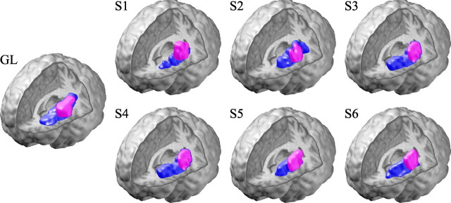
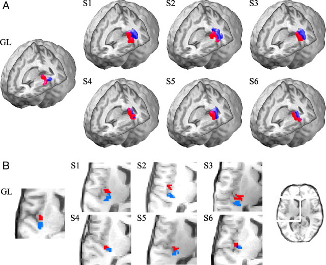
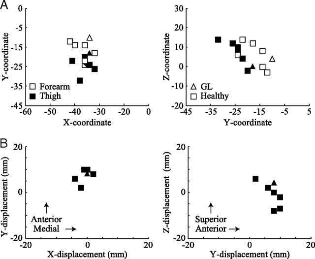
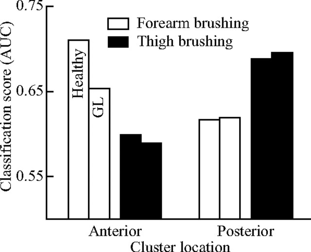
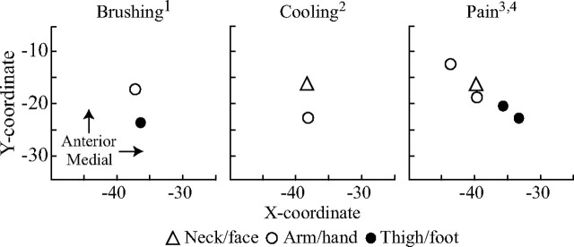
A network of thin (C and A delta) afferents relays various signals related to the physiological condition of the body, including sensations of gentle touch, pain, and temperature changes. Such afferents project to the insular cortex, where a somatotopic organization of responses to noxious and cooling stimuli was recently observed. To explore the possibility of a corresponding body-map topography in relation to gentle touch mediated through C tactile (CT) fibers, we applied soft brush stimuli to the right forearm and thigh of a patient (GL) lacking A beta afferents, and six healthy subjects during functional magnetic resonance imaging (fMRI). For improved fMRI analysis, we used a highly sensitive multivariate voxel clustering approach. A somatotopic organization of the left (contralateral) posterior insular cortex was consistently demonstrated in all subjects, including GL, with forearm projecting anterior to thigh stimulation. Also, despite denying any sense of touch in daily life, GL correctly localized 97% of the stimuli to the forearm or thigh in a forced-choice paradigm. The consistency in activation patterns across GL and the healthy subjects suggests that the identified organization reflects the central projection of CT fibers. Moreover, substantial similarities of the presently observed insular activation with that described for noxious and cooling stimuli solidify the hypothesized sensory-affective role of the CT system in the maintenance of physical well-being as part of a thin-afferent homeostatic network.


Figures

Figure 1.
Figure 1.
Insular region of interest. Voxel clusters maximally differentiating a soft brush stimulation from rest (purple) in the neuronopathy syndrome patient GL (lacking thick, myelinated afferents) and six healthy subjects (S1–S6). The brushing stimulation analysis was restricted to the manually identified contralateral (left) insular cortex (blue). Note that the response is consistently located to the posterior insular cortex across all subjects, including GL, in line with our previous observations (Olausson et al., 2002, 2008).
Figure 2.
Figure 2.
Insular somatotopy in response to gentle touch. A, The posterior contralateral (left) insular regions maximally differentiating forearm (red) and thigh (blue) soft brush stimulation from rest in the neuronopathy syndrome patient GL and six healthy subjects (S1–S6). Cluster overlap is shown in dark blue. The somatotopic organization was consistently identified in all subjects, with forearm brushing projecting anterior to thigh brushing. B, Horizontal slices (MNI Z = 6) of the same clusters, highlighting the anterior–posterior distinction.
Figure 3.
Figure 3.
Forearm and thigh projections in response to gentle touch. A, B, The forearm (white) and thigh (black) cluster centroid MNI coordinates (A) and displacements (B; forearm relative to thigh) for each of the healthy subjects (squares) and the neuronopathy syndrome patient GL (triangles) clearly illustrate the observed somatotopic organization of the contralateral (left) posterior insular cortex. The healthy subjects showed a significant difference between forearm and thigh cluster centroid location in the Y-plane only, with forearm located anterior to thigh stimulation (two-tailed paired t test, p < 0.05).
Figure 4.
Figure 4.
Cluster selectivity verification. The classification scores (measured by AUC, where a value of 1 equals perfect separation) of the forearm (white) and thigh (black) brushing volumes on the posterior insular voxels obtained using the clustering algorithm (as seen in Fig. 2). The healthy subject (left bar) forearm brushing volumes were significantly more separable in the forearm cluster than in the thigh cluster, and vice versa for the thigh brushing volumes (two-tailed paired t test, p < 0.05). Also, the forearm brushing scores were significantly higher than those of thigh brushing in the anterior cluster, and vice versa (two-tailed paired t test, p < 0.05). The neuronopathy patient data (GL; right bar) follow the healthy subject trend. These results verify the forearm–anterior and thigh–posterior pattern demonstrated by the clustering algorithm.
Figure 5.
Figure 5.
Thin-fiber activation center comparison. Posterior insular cortex activation centers (in MNI coordinates) resulting from application of thin-fiber exciting stimuli (soft brushing, innocuous cooling, and pain) on various body parts reported in the present1 and previous studies (Hua et al., 20052, Henderson et al., 20073; Brooks et al., 20054). White markers indicate an upper body stimulus location and black a lower body location. Gentle tactile stimulation (brushing) fit the somatotopic pattern in the posterior contralateral insular cortex well, with upper body stimulations projecting anterior (and slightly lateral) to lower body stimulations. The nature of the relative spatial organization of these modalities within the posterior insular cortex has yet to be demonstrated in humans, however.

Similar articles

Cited by

References

    1. Bandler R, Price JL, Keay KA. Brain mediation of active and passive emotional coping. Prog Brain Res. 2000;122:333–349. - PubMed
    1. Bessou P, Burgess PR, Perl ER, Taylor CB. Dynamic properties of mechanoreceptors with unmyelinated (C) fibers. J Neurophysiol. 1971;34:116–131. - PubMed
    1. Björnsdotter Åberg M, Wessberg J. An evolutionary approach to the identification of informative voxel clusters for brain state discrimination. IEEE J Select Topics Signal Proc. 2008;2:919–928.
    1. Blomqvist A, Craig AD. Is neuropathic pain caused by the activation of nociceptive-specific neurons due to anatomic sprouting in the dorsal horn? J Comp Neurol. 2000;428:1–4. - PubMed
    1. Brooks JC, Zambreanu L, Godinez A, Craig AD, Tracey I. Somatotopic organisation of the human insula to painful heat studied with high resolution functional imaging. Neuroimage. 2005;27:201–209. - PubMed

Publication types

MeSH terms