. 2009 Jul 22;29(29):9330-43.
doi: 10.1523/JNEUROSCI.2212-09.2009.

Extrasynaptic NMDA receptors couple preferentially to excitotoxicity via calpain-mediated cleavage of STEP

Affiliations

Extrasynaptic NMDA receptors couple preferentially to excitotoxicity via calpain-mediated cleavage of STEP

Jian Xu et al. J Neurosci. .

Abstract

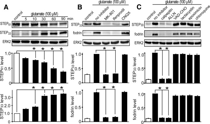
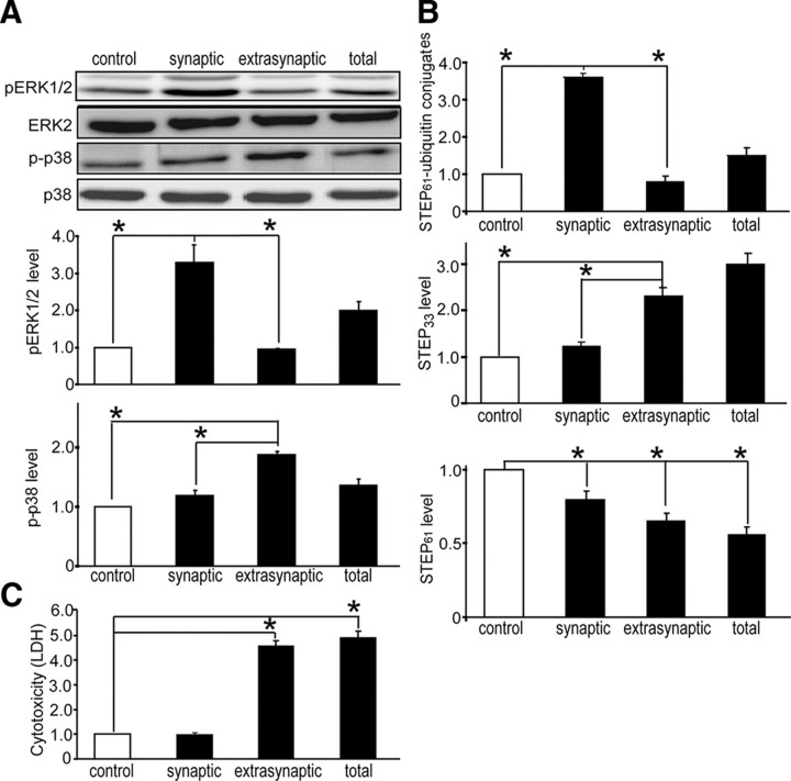
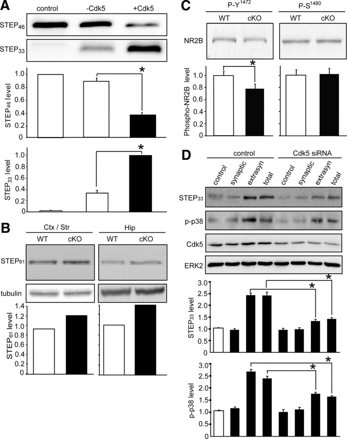
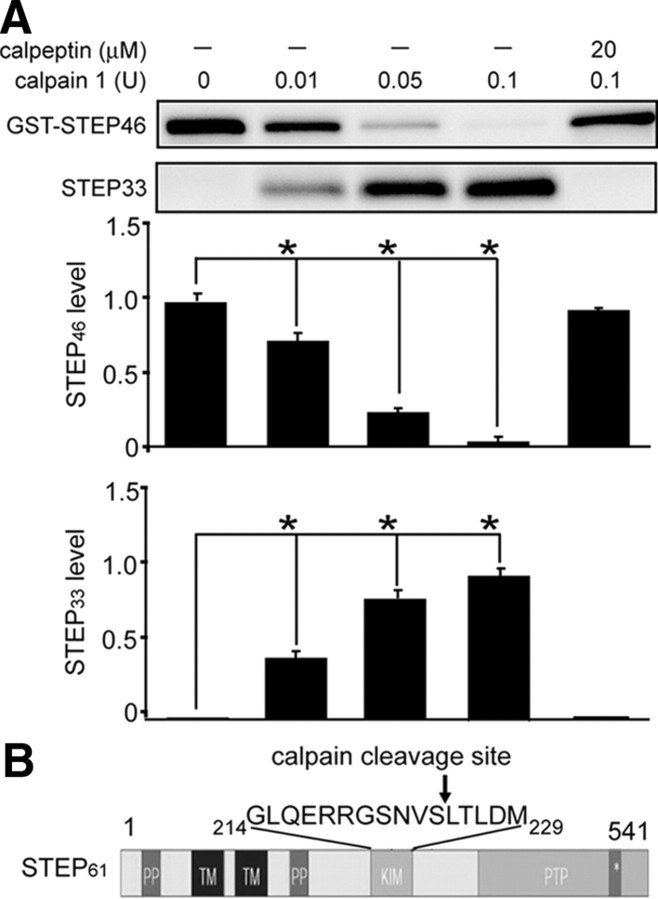
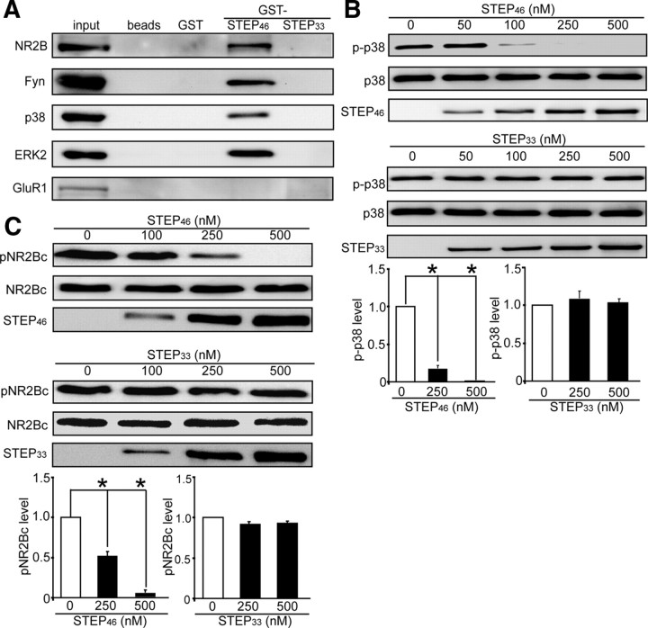
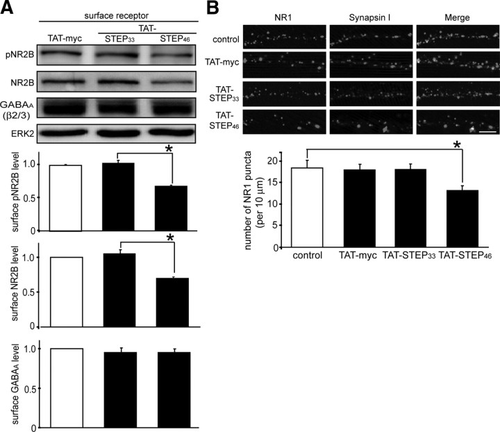
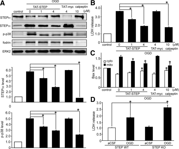
NMDA receptor (NMDAR)-mediated excitotoxicity plays an important role in several CNS disorders, including epilepsy, stroke, and ischemia. Here we demonstrate the involvement of striatal-enriched protein tyrosine phosphatase (STEP) in this critical process. STEP(61) is an alternatively spliced member of the family that is present in postsynaptic terminals. In an apparent paradox, STEP(61) regulates extracellular signal-regulated kinase 1/2 (ERK1/2) and p38, two proteins with opposing functions; activated p38 promotes cell death, whereas activated ERK1/2 promotes cell survival. We found that synaptic stimulation of NMDARs promoted STEP(61) ubiquitination and degradation, concomitant with ERK1/2 activation. In contrast, extrasynaptic stimulation of NMDARs invoked calpain-mediated proteolysis of STEP(61), producing the truncated cleavage product STEP(33) and activation of p38. The calpain cleavage site on STEP was mapped to the kinase interacting motif, a domain required for substrate binding. As a result, STEP(33) neither interacts with nor dephosphorylates STEP substrates. A synthetic peptide spanning the calpain cleavage site efficiently reduced STEP(61) degradation and attenuated p38 activation and cell death in slice models. Furthermore, this peptide was neuroprotective when neurons were subjected to excitotoxicity or cortical slices were exposed to ischemic conditions. These findings suggest a novel mechanism by which differential NMDAR stimulation regulates STEP(61) to promote either ERK1/2 or p38 activation and identifies calpain cleavage of STEP(61) as a valid target for the development of neuroprotective therapy.


Figures

Figure 1.
Figure 1.
NMDAR-dependent cleavage of STEP61 by calpain. A, Immunoblots of STEP61 and STEP33 from primary cortical neurons lysates treated with glutamate for the indicated periods. Representative immunoblots (top) and quantitation (bottom) are shown for each (*p < 0.01 compared with control, one-way ANOVA with post hoc Tukey's test; n = 3). ERK2 was used to normalize the data. B, Immunoblots of STEP33 and fodrin from cortical neurons treated with glutamate in the presence of MK-801 (50 μm), ifenprodil (10 μm), or CNQX (40 μm). Activation of calpain was confirmed by fodrin proteolysis. Histogram shows the quantitative analysis of STEP33 production and fodrin cleavage normalized to ERK2 (*p < 0.01, Student's t test; n = 3). C, Immunoblot analyses are shown of lysates from cortical neurons treated with glutamate in the presence of the calpain inhibitors calpeptin (10 μm) or ALLN (20 μm), the caspase inhibitor cpm-VAD-CHO (20 μm), the proteasome inhibitors lactacystin (5 μm) or epoximicin (5 μm), or the lyzosome inhibitor chloroquine (500 μm) (*p < 0.01, Student's t test; n = 3).
Figure 2.
Figure 2.
Activation of extrasynaptic NMDARs causes STEP61 cleavage and neuronal death. A, Immunoblot analysis comparing the effects of synaptic or extrasynaptic NMDAR stimulation on the phosphorylation of ERK1/2 and p38. Quantitative analyses for each were normalized to ERK2 and are summarized in the bar graphs (*p < 0.01, one-way ANOVA with post hoc Tukey's test; n = 4). B, Synaptic stimulation in the presence of proteasome inhibitor MG-132 led to accumulation of STEP61–ubiquitin conjugates (top), whereas extrasynaptic stimulation led to production of STEP33 (middle). Both synaptic and extrasynaptic stimulation of NMDARs led to a decrease in STEP61 levels (bottom). ERK2 was used to normalize the data (*p < 0.01, one-way ANOVA with post hoc Tukey's test; n = 4). C, Analysis of excitotoxic cell death of cortical neurons subjected to synaptic or extrasynaptic stimulations. Cytotoxicity was assessed by measurement of LDH release (*p < 0.01, Student's t test; n = 3).
Figure 3.
Figure 3.
Regulation of STEP cleavage by Cdk5. A, GST–STEP46 was digested with calpain 1 in the absence or presence of recombinant Cdk5/p25 proteins (50 ng/μl). Immunoreactivity of noncleaved GST–STEP46 was normalized to control without calpain (*p < 0.01, one-way ANOVA with post hoc Tukey's test; n = 3). B, Immunoblot analysis and quantitation of STEP61 and tubulin loading controls in corticostriatal and hippocampal synaptosomes. Tissues from three animals per genotype were pooled. Ctx, Cortex; Str, striatum; Hip, hippocampus. C, Levels of indicated phosphorylation sites in hippocampal lysates from Cdk5 cKO and WT mice. Phosphorylation sites are normalized to total protein levels (*p < 0.05, Student's t test; n = 6). D, STEP cleavage and p38 activation in Cdk5 siRNA-transfected cortical neurons. Extrasynaptic stimulation and total glutamate bath-induced STEP cleavage was significantly reduced in Cdk5 siRNA-transfected neurons. Moreover, activation of p38 was significantly blocked in Cdk5 siRNA-transfected neurons during extrasynaptic stimulation as well as total glutamate bath (*p < 0.01, one-way ANOVA with post hoc Tukey's test; n = 3).
Figure 4.
Figure 4.
Identification of calpain cleavage of site. A, Recombinant GST–STEP46 was digested with calpain 1 at the indicated concentrations in the absence or presence of calpeptin (20 μm) (*p < 0.01, one-way ANOVA with post hoc Tukey's test; n = 3). B, After calpain digestion, the C-terminal fragment (STEP33) was sent for amino acid sequencing. Sequence is shown surrounding the cleavage site within the KIM domain. PP, Polyproline-rich domain; TM, transmembrane; PTP, protein tyrosine phosphatase domain.
Figure 5.
Figure 5.
STEP33 does not interact with STEP substrates. A, The substrate-trapping (C to S) mutant of STEP46 or STEP33 (10 μg) were coupled to glutathione Sepharose beads and incubated with rat brain homogenates (100 μg). Bound proteins were immunoblotted with indicated antibodies. GST protein and glutathione Sepharose beads were used as negative controls. Representative blot was shown from three independent experiments. B, p-p38 (10 ng) was incubated with STEP33 or STEP46 at the indicated concentrations and blotted with phospho-p38, total p38, or STEP antibodies. p-p38 levels were normalized to total p38 (*p < 0.01, one-way ANOVA with post hoc Tukey's test; n = 3). C, GST–NR2Bc was phosphorylated by active Fyn, incubated with STEP33 or STEP46 at the indicated concentrations, and blotted with pNR2B (pY1472), total NR2B, or STEP antibodies. pNR2B levels were normalized to total NR2B (*p < 0.01, one-way ANOVA with post hoc Tukey's test; n = 3).
Figure 6.
Figure 6.
STEP33 blocks NMDAR endocytosis. A, Corticostriatal slices were treated with TAT–myc, TAT–STEP33, or TAT–STEP46 (2 μm) and processed to obtain synaptic membrane fractions (LP1). Phospho-specific and total NR2B subunits were analyzed with pY1472 NR2B (pNR2B) and NR2B antibodies, respectively. Expression of GABAA (β2/3) was used as a control. All blots were normalized to total ERK2 levels. Bar graphs show the quantitative analysis of each receptor (*p < 0.01, one-way ANOVA with post hoc Tukey's test; n = 3). B, Cortical neurons were treated with TAT–myc, TAT–STEP33, or TAT–STEP46 (2 μm) for 30 min, followed by immunostaining with anti-NR1 (green) and anti-synapsin I (red) antibodies. Merged images are also shown. The number of NR1 puncta was counted per 10 μm of three or more dendrites per neuron, and 20 neurons were used for quantification per treatment and are shown in the bar graph below (*p < 0.01, one-way ANOVA with post hoc Tukey's test; n = 20). Scale bar, 5 μm.
Figure 7.
Figure 7.
TAT–STEP peptide is neuroprotective against glutamate excitotoxicity. A, GST–STEP46 was digested with calpain 1 in the presence of TAT–STEP peptide at the indicated concentrations. Calpeptin (10 μm) served as a control. Uncleaved STEP was normalized to no treatment control (*p < 0.01, one-way ANOVA with post hoc Tukey's test; n = 3). B, Cortical neurons were pretreated with TAT–STEP peptide or calpeptin (10 μm), followed by glutamate stimulation for 1 h. Total cell lysates were blotted with antibodies against STEP and ERK2. Production of STEP33 was normalized to total ERK2 levels (*p < 0.01, one-way ANOVA with post hoc Tukey's test; n = 3). C, Cell cytotoxicity was assessed by LDH release after synaptic or extrasynaptic stimulations. TAT–STEP peptide, TAT–myc, or calpeptin was preincubated as indicated before extrasynaptic stimulation (*p < 0.01, Student's t test; n = 3). D, Cortical cultures were treated as in C, and cell viability was measured by MAP2 staining (*p < 0.01, Student's t test; n = 3). E, Cell viability was quantified using DAPI staining. Arrows indicate injured or dead neurons, which show nuclear morphological changes (e.g., chromatin condensation, nuclear condensation, and segmentation). At least 250 randomly selected cells were counted for each stimulation (*p < 0.01, Student's t test). Scale bar, 10 μm.
Figure 8.
Figure 8.
TAT–STEP peptide is neuroprotective in the OGD model. A, Top, Corticostriatal slices were subjected to OGD for 2 h, followed by 2 h reoxygenation in the absence or presence of TAT–STEP peptide, TAT–myc, or calpeptin. At the end of treatment, slices processed by immunoblotting with antibodies against STEP, fodrin, p-p38, or ERK2. Quantification of production of STEP33 (*p < 0.01, one-way ANOVA with post hoc Tukey's test; n = 3) after normalization to total ERK2 levels (middle). Quantification of phospho-p38 is shown in the bottom (*p < 0.01, one-way ANOVA with post hoc Tukey's test; n = 3). B, LDH release was measured at the end of treatment (*p < 0.01, one-way ANOVA with post hoc Tukey's test; n = 3). C, Quantification of Bax in cytosol and mitochondrial fractions after normalization to tubulin (cytosol) or COX IV (mitochondria) (*p < 0.01, one-way ANOVA with post hoc Tukey's test; n = 3). D, Corticostriatal slices from STEP WT or KO mice were subjected to OGD stimulation. Bar graph shows the quantitative analysis of LDH release from each group (*p < 0.01, one-way ANOVA with post hoc Tukey's test; n = 10).

Similar articles

Cited by

References

    1. Aarts M, Liu Y, Liu L, Besshoh S, Arundine M, Gurd JW, Wang YT, Salter MW, Tymianski M. Treatment of ischemic brain damage by perturbing NMDA receptor- PSD-95 protein interactions. Science. 2002;298:846–850. - PubMed
    1. Arundine M, Tymianski M. Molecular mechanisms of calcium-dependent neurodegeneration in excitotoxicity. Cell Calcium. 2003;34:325–337. - PubMed
    1. Bano D, Young KW, Guerin CJ, Lefeuvre R, Rothwell NJ, Naldini L, Rizzuto R, Carafoli E, Nicotera P. Cleavage of the plasma membrane Na+/Ca2+ exchanger in excitotoxicity. Cell. 2005;120:275–285. - PubMed
    1. Besshoh S, Bawa D, Teves L, Wallace MC, Gurd JW. Increased phosphorylation and redistribution of NMDA receptors between synaptic lipid rafts and post-synaptic densities following transient global ischemia in the rat brain. J Neurochem. 2005;93:186–194. - PubMed
    1. Boland B, Campbell V. Beta-amyloid (1–40)-induced apoptosis of cultured cortical neurones involves calpain-mediated cleavage of poly-ADP-ribose polymerase. Neurobiol Aging. 2003;24:179–186. - PubMed

Publication types

MeSH terms

LinkOut - more resources