. 2009 Sep;102(3):1994-2006.
doi: 10.1152/jn.00613.2009. Epub 2009 Jul 22.

Alternative splicing in the voltage-gated sodium channel DmNav regulates activation, inactivation, and persistent current

Affiliations

Alternative splicing in the voltage-gated sodium channel DmNav regulates activation, inactivation, and persistent current

Wei-Hsiang Lin et al. J Neurophysiol. 2009 Sep.

Abstract

Diversity in neuronal signaling is a product not only of differential gene expression, but also of alternative splicing. However, although recognized, the precise contribution of alternative splicing in ion channel transcripts to channel kinetics remains poorly understood. Invertebrates, with their smaller genomes, offer attractive models to examine the contribution of splicing to neuronal function. In this study we report the sequencing and biophysical characterization of alternative splice variants of the sole voltage-gated Na+ gene (DmNav, paralytic), in late-stage embryos of Drosophila melanogaster. We identify 27 unique splice variants, based on the presence of 15 alternative exons. Heterologous expression, in Xenopus oocytes, shows that alternative exons j, e, and f primarily influence activation kinetics: when present, exon f confers a hyperpolarizing shift in half-activation voltage (V1/2), whereas j and e result in a depolarizing shift. The presence of exon h is sufficient to produce a depolarizing shift in the V1/2 of steady-state inactivation. The magnitude of the persistent Na+ current, but not the fast-inactivating current, in both oocytes and Drosophila motoneurons in vivo is directly influenced by the presence of either one of a pair of mutually exclusive, membrane-spanning exons, termed k and L. Transcripts containing k have significantly smaller persistent currents compared with those containing L. Finally, we show that transcripts lacking all cytoplasmic alternatively spliced exons still produce functional channels, indicating that splicing may influence channel kinetics not only through change to protein structure, but also by allowing differential modification (i.e., phosphorylation, binding of cofactors, etc.). Our results provide a functional basis for understanding how alternative splicing of a voltage-gated Na+ channel results in diversity in neuronal signaling.


Figures

FIG. 1.
FIG. 1.
Characterization of 27 open reading frames (ORFs) of DmNav isolated from late-stage 17 embryos. A: schematic of the predicted 4-homology domain (I, II, III, and IV) structure of DmNav protein with approximate locations of spliced exons shown. Exons j, 7, 8, i, a, b, e, f, 22, h, 23 are optional, whereas exons c/d and k/L are mutually exclusive. B: exon usage of the 27 individual clones isolated. The variant number (DmNavX) and frequency of clones are indicated. The clones in the shaded box (i.e., 1, 26, and 28) are also found in adult heads (O'Donnell Olson et al. 2008).
FIG. 2.
FIG. 2.
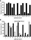
Frequency of alternative exon usage. A: analysis of exon usage across all 27 DmNav ORFs shows that exons 7, 8, i, b, d, f, 22, 23, and L are significantly more common (present in >75% of clones) than j, c, e, h, and k. Exon a exhibits a more variable frequency of inclusion, being present in 60% of clones. B: comparison of exon frequency in embryo and adult CNS for those exons that have been analyzed at both developmental stages. The data for adults are taken from O'Donnell Olson et al. (2008).
FIG. 3.
FIG. 3.
Voltage dependence of activation is affected by inclusion of exons f, j, and e. Activation curves (means ± SE for n ≥ 8) are shown for 3 paired comparisons (DmNav31 vs. 10, 34 vs. 10h, and 26 vs. 10k) in which identical clones differ by either containing f+, j, e, or f, j+, e+. Inclusion of exon f and loss of exons j and e result in a hyperpolarizing shift in half-activation voltage (V1/2). See Table 3 for actual V1/2 values. All paired comparisons are significantly different at P ≤ 0.01. Example traces are shown on the left. Scale bars are 5 ms and 0.5 μA, respectively. Voltage dependence of activation was determined by 100-ms depolarizing potentials from −80 to +5 mV (in 5-mV steps) applied from a holding prepulse potential of −90 mV.
FIG. 4.
FIG. 4.
Voltage dependence of steady-state inactivation is influenced by inclusion of exon h. Inactivation curves (means ± SE for n ≥ 8) are shown for 3 paired comparisons (DmNav31 vs. 34, 30 vs. 32, and 10 vs. 10h), in which identical clones differ by either containing h+ or h. Inclusion of exon h results in a depolarizing shift in V1/2 of inactivation. See Table 4 for actual V1/2 values. All paired comparisons are significantly different at P ≤ 0.05. Example traces are shown on the left. Scale bars are 5 ms and 0.5 μA, respectively. Voltage dependence of steady-state inactivation was determined by 100-ms depolarizing prepotentials from −80 to +5 mV (in 5-mVsteps) followed by a test potential of −10 mV for 50 ms.
FIG. 5.
FIG. 5.
Amount of persistent current is influenced by exons k and L. Measurement of the percentage of persistent (P) current relative to transient current (T) in 3 paired comparisons (DmNav 26 vs. 31, 10k vs. 10, and 49k vs. 49), in which identical clones differ by either containing k+, L or k, L+. Inclusion of exon L results in a marked increase in persistent relative to transient current. Actual values are listed in Table 5. Example traces are shown on the left and averaged values (%) of persistent current (means ± SE for n ≥ 8) on the right (P < 0.01 for all comparisons). For ease of analysis, all example traces have been scaled to have the same amplitude of transient current. Scale bars are 5 ms and 0.5 μA, respectively. Voltage commands were the same as those for activation voltage determination (see Fig. 3). Persistent current was measured at the end of a depolarizing command voltage. The largest persistent current was normalized by division with the largest transient current obtained (i.e., not necessarily from the same voltage step).
FIG. 6.
FIG. 6.
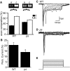
The presence of exon k reduces persistent Na+ current in Drosophila motoneurons. A: comparison of expression of DmNav transcripts containing either exon k or L in both wild-type (WT, Canton-S) and pasilla (ps) mutant CNS in late-stage 17 embryos. Semiquantitative polymerase chain reaction (PCR) shows that, in WT, expression of L-containing DmNav predominates (66.8 ± 0.9%) relative to inclusion of exon k (33.2 vs. 0.9%). By contrast, in the absence of ps, exon frequencies are more similar (54.0 ± 0.8 vs. 46.0 ± 0.8%, L vs. k, respectively). The differences in exon composition between WT and ps are significant at P < 0.01. Example images of PCR products are shown for both WT and ps (images have been manipulated using Photoshop) and the average of 5 independent PCR experiments are shown (means ± SE). B: whole cell patch recordings from identified motoneurons (aCC and RP2) show that the percentage persistent Na+ current, relative to the transient component, is significantly reduced in the absence of ps (20.5 ± 2.4 vs. 14.0 ± 1.5%, WT vs. ps, respectively; P ≤ 0.05, n ≥ 10, mean ± SE). Persistent current was measured at the end of a depolarizing command voltage. The largest persistent current was normalized by division with the largest transient current obtained (i.e., not necessarily from the same voltage step). C and D: example whole cell traces of Na+ currents obtained from aCC/RP2 motoneurons in both WT (C) and ps (D) late-stage 17 embryos. The 2 traces were chosen because the peak transient amplitude was the same in both recordings (i.e., the 2 traces are not scaled). E: voltage steps (50 ms) from −60 to +20 mV (in 10-mV steps) were applied from a holding prepulse potential of −90 mV.
FIG. 7.
FIG. 7.
Cytoplasmic spliced exons are not required for channel function. Removal of all cytoplasmic spliced exons (j, i, a, b, e, f, and h) does not abolish channel function, indicating that these exons are required to modify only gating properties. The minimal clone expressed contains only spliced exons d and L. Ai and Aii: comparison of DmNaVminimal with that of the most common embryonic (DmNaV30) and adult (DmNaV1) splice variant shows removal of all cytoplasmic spliced exons results in a significant hyperpolarization of V1/2 activation (see Table 3 for precise values). Bi and Bii: DmNaVminimal also exhibits a significantly depolarized V1/2 inactivation compared with either DmNaV1 or 30. Ci and Cii: DmNaVminimal exhibits the greatest degree of persistent current compared with either DmNaV1 or 30. Example traces for activation and inactivation are shown on the left and averaged values (means ± SE for n ≥ 8) on the right (DmNaVminimal is different from either DmNaV1 or 30 at P < 0.05 for all comparisons).
FIG. 8.
FIG. 8.
DmNav exons k and L are conserved in human voltage-gated Na+ channels. Mutually exclusive DmNav spliced exons k and L differ by 16/41 residues (shown in gray boxes in the exon k sequence). Analysis of exon 18 of human Nav1.1–1.9 shows that it is more similar to DmNav exon L (identical residues shown in black) than to exon k.

Similar articles

Cited by

References

    1. Auld VJ, Goldin AL, Krafte DS, Marshall J, Dunn JM, Catterall WA, Lester HA, Davidson N, Dunn RJ. A rat brain Na+ channel alpha subunit with novel gating properties. Neuron 1: 449–461, 1988. - PubMed
    1. Baines RA. Postsynaptic protein kinase A reduces neuronal excitability in response to increased synaptic excitation in the Drosophila CNS. J Neurosci 23: 8664–8672, 2003. - PMC - PubMed
    1. Baines RA, Bate M. Electrophysiological development of central neurons in the Drosophila embryo. J Neurosci 18: 4673–4683, 1998. - PMC - PubMed
    1. Belcher SM, Zerillo CA, Levenson R, Ritchie JM, Howe JR. Cloning of a sodium channel alpha subunit from rabbit Schwann cells. Proc Natl Acad Sci USA 92: 11034–11038, 1995. - PMC - PubMed
    1. Brose K, Tessier-Lavigne M. Slit proteins: key regulators of axon guidance, axonal branching, and cell migration. Curr Opin Neurobiol 10: 95–102, 2000. - PubMed

Publication types

LinkOut - more resources