. 2009 Aug 4;106(31):13004-9.
doi: 10.1073/pnas.0906387106. Epub 2009 Jul 22.

Preerythrocytic, live-attenuated Plasmodium falciparum vaccine candidates by design

Affiliations

Preerythrocytic, live-attenuated Plasmodium falciparum vaccine candidates by design

Kelley M VanBuskirk et al. Proc Natl Acad Sci U S A. .

Abstract

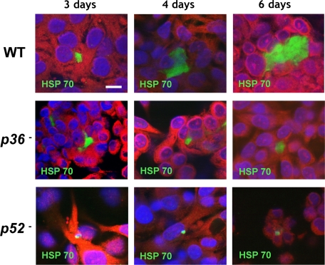
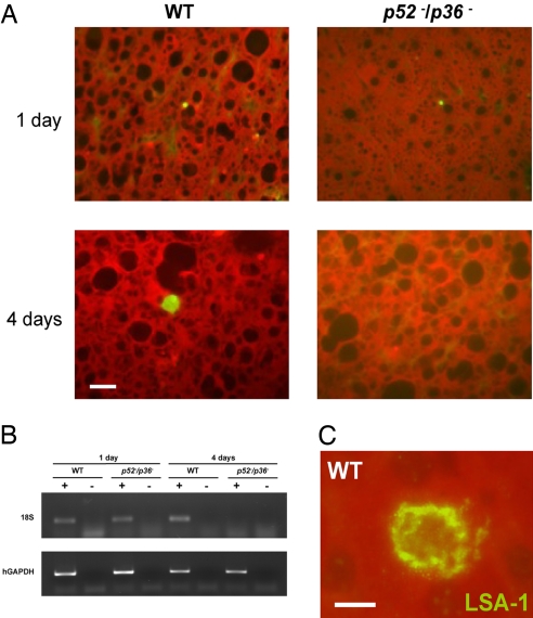
Falciparum malaria is initiated when Anopheles mosquitoes transmit the Plasmodium sporozoite stage during a blood meal. Irradiated sporozoites confer sterile protection against subsequent malaria infection in animal models and humans. This level of protection is unmatched by current recombinant malaria vaccines. However, the live-attenuated vaccine approach faces formidable obstacles, including development of accurate, reproducible attenuation techniques. We tested whether Plasmodium falciparum could be attenuated at the early liver stage by genetic engineering. The P. falciparum genetically attenuated parasites (GAPs) harbor individual deletions or simultaneous deletions of the sporozoite-expressed genes P52 and P36. Gene deletions were done by double-cross-over recombination to avoid genetic reversion of the knockout parasites. The gene deletions did not affect parasite replication throughout the erythrocytic cycle, gametocyte production, mosquito infections, and sporozoite production rates. However, the deletions caused parasite developmental arrest during hepatocyte infection. The double-gene deletion line exhibited a more severe intrahepatocytic growth defect compared with the single-gene deletion lines, and it did not persist. This defect was assessed in an in vitro liver-stage growth assay and in a chimeric mouse model harboring human hepatocytes. The strong phenotype of the double knockout GAP justifies its human testing as a whole-organism vaccine candidate using the established sporozoite challenge model. GAPs might provide a safe and reproducible platform to develop an efficacious whole-cell malaria vaccine that prevents infection at the preerythrocytic stage.


Conflict of interest statement

Conflict of interest statement: S.H.I.K. is an inventor listed on U.S. Patent No. 7,22,179, U.S. Patent No. 7,261,884, and international patent application PCT/US2004/043023, each titled “Live Genetically Attenuated Malaria Vaccine.”

Figures

Fig. 1.
Fig. 1.
The p52- and p36-deficient P. falciparum parasites show defective liver-stage development. Development of mutant and WT liver stages was assessed in vitro by using cultured cells of the HC-04 human hepatocytic line. Parasite growth was monitored over 6 days, and liver stages were visualized by immunofluorescence microscopy at 400× magnification with anti-HSP70 at 3, 4, and 6 days after infection. P. falciparum p52 and p36 parasites exhibited abnormal, arrested development that is most apparent at the 6-day developmental time point. (Scale bar: 10 μm.)
Fig. 2.
Fig. 2.
Phenotypic analysis of P. falciparum p52/p36-deficient sporozoites and liver stages reveals a severe defect in liver-stage development. (A) The p52/p36 parasites show normal invasion of the mosquito salivary glands; no significant differences were observed between WT and the double-knockout clones (P = 0.89). (B) The p52/p36 parasites showed slightly lower gliding activity compared with WT parasites; the effect was not statistically significant at the 95% confidence level (P = 0.11). (C) WT and p52/p36 parasites have comparable ability to enter hepatocytes; no significant difference was seen between p52/p36 and WT parasites (P = 0.11). (D) The double-knockout parasites show a severe developmental arrest and do not persist, because no p52/p36 liver stages are detected at 4 days after HC-04 cell line invasion. Statistical differences between the mutant and WT parasite lines were evaluated by the Wilcoxon matched-pairs, signed-rank test. nd, not detected.
Fig. 3.
Fig. 3.
P. falciparum p52/p36 parasites do not grow and persist in a humanized chimeric liver mouse model. SCID Alb-uPA mice were inoculated with 106 primary human hepatocytes to create chimeric human–mouse livers. Mice positive for engraftment were infected intravenously with 1 × 10⁶ sporozoites of either p52/p36 or WT P. falciparum. (A) Immunofluorescent micrographs of chimeric SCID/Alb-uPA liver cryosections after WT P. falciparum infection, stained with parasite-specific antibodies. Tissue was harvested at 1 day or 4 days after mice were inoculated with P. falciparum sporozoites. Sections in panels were stained with an anti-PfHSP70 monoclonal antibody. The data show that p52/p36 liver stages are detected 1 day after infection but are not detected 4 days after infection. (Scale bar: 10 μm.) (B) RT-PCR analysis was performed with RNA isolated from individual livers of chimeric mice 1 day or 4 days after infection. Total RNA was extracted, and reverse-transcription reactions were done with (+) or without (−) the reverse transcriptase. Primers specific for the 18S (small-subunit) ribosomal RNA of P. falciparum and human glyceraldehyde phosphate dehydrogenase (hGAPDH) were then used to amplify the reverse-transcribed message. The GAPDH control reaction was positive for samples from all of the mice. The parasite-specific 18S reaction was positive for the mice infected with the WT parasites at day 1 and day 4 after infection. For the double-knockout parasite-infected mice, the 18S reaction was only positive for tissues harvested 1 day after infection and negative for tissue harvested at 4 days after infection, indicating that p52/p36 parasites were no longer present in the liver at the later time point. RT-PCR was performed on all mice used in the experiment, and results were consistent with the results shown here. (C) An example of WT P. falciparum liver stage from chimeric SCID/Alb-uPA liver cryosection stained with polyclonal rabbit antisera against the repeat region of LSA-1 at 4 days after infection. (Scale bar: 5 μm.)

Similar articles

Cited by

References

    1. Snow RW, Guerra CA, Noor AM, Myint HY, Hay SI. The global distribution of clinical episodes of Plasmodium falciparum malaria. Nature. 2005;434:214–217. - PMC - PubMed
    1. Nussenzweig RS, Vanderberg J, Most H, Orton C. Protective immunity produced by the injection of x-irradiated sporozoites of plasmodium berghei. Nature. 1967;216:160–162. - PubMed
    1. Clyde DF, Most H, McCarthy VC, Vanderberg JP. Immunization of man against sporozite-induced falciparum malaria. Am J Med Sci. 1973;266:169–177. - PubMed
    1. Rieckmann KH, Carson PE, Beaudoin RL, Cassells JS, Sell KW. Letter: Sporozoite induced immunity in man against an Ethiopian strain of Plasmodium falciparum. Trans R Soc Trop Med Hyg. 1974;68:258–259. - PubMed
    1. Hoffman SL, et al. Protection of humans against malaria by immunization with radiation-attenuated Plasmodium falciparum sporozoites. J Infect Dis. 2002;185:1155–1164. - PubMed

Publication types

LinkOut - more resources