. 2009 Aug 4;106(31):12932-7.
doi: 10.1073/pnas.0906606106. Epub 2009 Jul 22.

The AMPK agonist AICAR inhibits the growth of EGFRvIII-expressing glioblastomas by inhibiting lipogenesis

Affiliations

The AMPK agonist AICAR inhibits the growth of EGFRvIII-expressing glioblastomas by inhibiting lipogenesis

Deliang Guo et al. Proc Natl Acad Sci U S A. .

Abstract

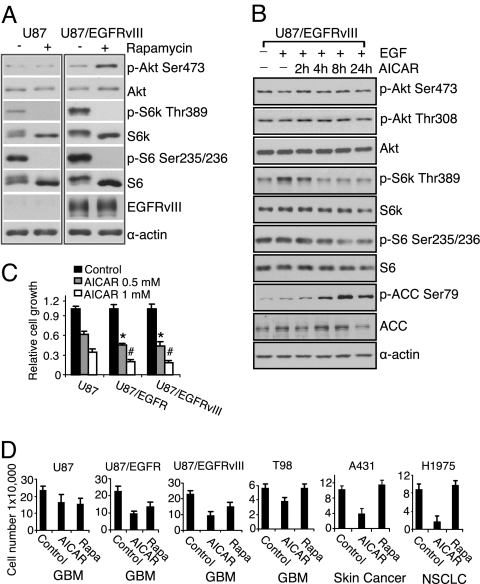
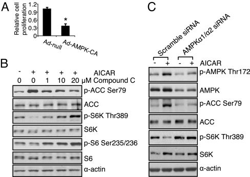
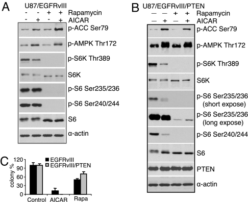
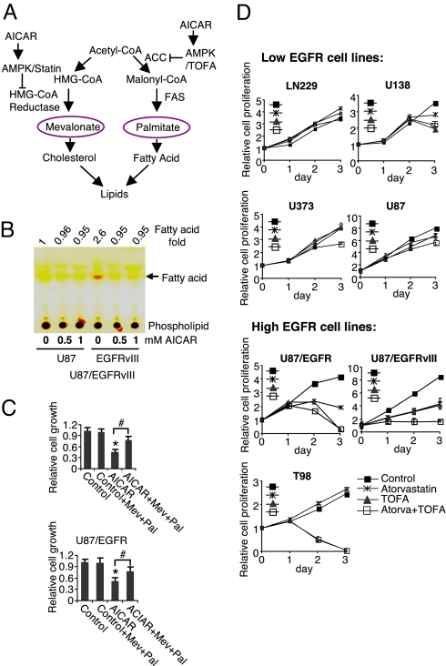
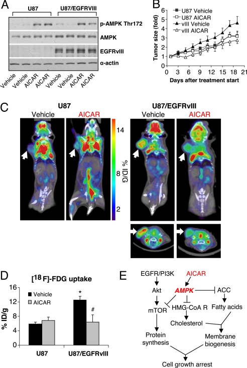
The EGFR/PI3K/Akt/mTOR signaling pathway is activated in many cancers including glioblastoma, yet mTOR inhibitors have largely failed to show efficacy in the clinic. Rapamycin promotes feedback activation of Akt in some patients, potentially underlying clinical resistance and raising the need for alternative approaches to block mTOR signaling. AMPK is a metabolic checkpoint that integrates growth factor signaling with cellular metabolism, in part by negatively regulating mTOR. We used pharmacological and genetic approaches to determine whether AMPK activation could block glioblastoma growth and cellular metabolism, and we examined the contribution of EGFR signaling in determining response in vitro and in vivo. The AMPK-agonist AICAR, and activated AMPK adenovirus, inhibited mTOR signaling and blocked the growth of glioblastoma cells expressing the activated EGFR mutant, EGFRvIII. Across a spectrum of EGFR-activated cancer cell lines, AICAR was more effective than rapamycin at blocking tumor cell proliferation, despite less efficient inhibition of mTORC1 signaling. Unexpectedly, addition of the metabolic products of cholesterol and fatty acid synthesis rescued the growth inhibitory effect of AICAR, whereas inhibition of these lipogenic enzymes mimicked AMPK activation, thus demonstrating that AMPK blocked tumor cell proliferation primarily through inhibition of cholesterol and fatty acid synthesis. Most importantly, AICAR treatment in mice significantly inhibited the growth and glycolysis (as measured by (18)fluoro-2-deoxyglucose microPET) of glioblastoma xenografts engineered to express EGFRvIII, but not their parental counterparts. These results suggest a mechanism by which AICAR inhibits the proliferation of EGFRvIII expressing glioblastomas and point toward a potential therapeutic strategy for targeting EGFR-activated cancers.


Conflict of interest statement

The authors declare no conflict of interest.

Figures

Fig. 1.
Fig. 1.
AICAR inhibits the growth of EGFR-activated cancer cell lines, including glioblastoma cells. (A) Western blot analysis demonstrated Akt Ser-473 phosphorylation in U87-EGFRvIII, but not U87 cells in response to rapamycin (1 nM) for 24 h. (B) Immunoblot analysis of effect of AICAR (0.5 mM) on Akt and mTORC1 signaling. U87-EGFRvIII cells were treated for up to 24 h and effect on signaling pathways was determined at indicated time points. AICAR (0.5 mM) treatment for up to 24 h inhibited mTOR signaling (S6K and S6 phosphorylation), but does not induce Akt phosphorylation. AICAR also phosphorylated AMPK downstream target gene ACC. (C) U87, U87-EGFRvIII, and U87-EGFR glioblastoma cells were seeded in 96-well plates for 24 h and then treated for 3 days with AICAR at the doses indicated. Relative growth was measured using the WST assay (Chemicon). AICAR was significantly more effective at blocking the growth of wild-type EGFR or EGFRvIII- expressing glioblastoma cells, relative to their parental U87 counterparts (*, P < 0.05; #, P < 0.05). (D) AICAR (0.5 mM) was more effective than rapamycin (Rapa 1 nM) in blocking the growth of cancer cells with activated EGFR signaling. Cell lines were treated with indicated drugs for 3 days, and cell number was determined by trypan exclusion. EGFR level of cell line is U87 (low expression), U87/EGFR (high expression), U87/EGFRvIII (high expression/mutation), T98 (high expression), A431 (amplification), and H1975 (L858R1/T790M mutation); cancer cell line type is indicated below the graph. Control cells were treated with ethanol, the diluent, alone (1:1,000).
Fig. 2.
Fig. 2.
The effect of AICAR on tumor growth and mTORC1 signaling is mediated through activation of AMPK. (A) Cells were infected by constitutively active AMPK adenovirus (Ad-AMPK-CA, 50 MOI) for 3 days, the growth of EGFRvIII expressing glioblastoma cells was inhibited by 65 ± 3.1%, *, P < 0.001 compared with cells infected by empty vector adenovirus (Ad-null, 50 MOI). (B) U87/EGFRvIII cells were treated with Compound C (1 μM, 10 μM, or 20 μM) for 30 min before AICAR (0.5 mM) treatment for 6 h. Cells were lysed and effect on signal transduction was determined by western blot using indicated antibodies. (C) U87/EGFRvIII cells were transfected with scrambled siRNA or AMPKα1/α2 siRNA (100 μM) for 48 h, then treated with AICAR (0.5 mM) for 6 h, followed by stimulation with EGF (20 ng/mL) for 15 min. Cellular lysates were the probed by western blot using indicated antibodies.
Fig. 3.
Fig. 3.
The anti-growth effect of AICAR is only partially mediated through inhibition of mTOR signaling. (A) U87/EGFRvIII cells were treated by AICAR (0.5 mM) or rapamycin (1 nM) for 6 h, then detected by western blotting using indicated antibodies. (B) Western blot analysis demonstrated that rapamycin, although less effective at inhibiting the growth of U87-EGFRvIII-PTEN cells, more completely inhibited S6K and S6 phorphorylation than did AICAR. (C) Reconstitution of PTEN desensitized U87-EGFRvIII glioblastoma cells to rapamycin, but not AICAR. Controls cells were treated by ethanol (1:1,000).
Fig. 4.
Fig. 4.
The anti-proliferative effect of AMPK activation is mediated by inhibition of lipogenesis. (A) Schema showing how AICAR can potentially regulate lipogenesis. (B) Thin layer chromatography of total cellular lipids from serum starved cells showed increased intracellular fatty acids in EGFRvIII expressing cells, which was inhibited by AICAR treatment for 24 h. (C) Addition of mevalonate and palmitate rescued AICAR-mediated growth inhibition. U87-EGFRvIII and U87-EGFR cells were plated, 24 h later they were treated with AICAR (0.5 mM) alone or in combination with mevalonate and/or palmitate (100 μM). Cells were treated for 3 days and relative cell growth was determined by the WST assay (Chemicon). *, P < 0.001 compared with control; # P < 0.01. (D) Inhibition of HMG-CoA reductase (atorvastatin 1 μM) and acetyl-CoA carboxylase (ACC) (TOFA 10 μg/mL) preferentially blocked the growth of EGFR-activated glioblastoma cells. Control cells were treated with DMSO (1:1,000).
Fig. 5.
Fig. 5.
AICAR blocks the growth of EGFRvIII expressing glioblastoma in vivo. (A) Western blot analysis of tumor xenograft lysates treated with AICAR (400 mg/kg) I.P. treatment for 3 days, or control (PBS only I.P. treatment), demonstrated AMPK Thr-172 phosphorylation in AICAR treated xenografts, but not control treated tumors. (B) AICAR significantly inhibited the growth of U87-EGFRvIII, but not U87 xenograft tumors, up to 21 days after initiation of treatment. Control mice were treated by PBS. (C and D) 18F-FDG microPET/CT imaging demonstrated that EGFRvIII expressing tumors have higher baseline (day 3) uptake of 18F-FDG relative to U87 cells; AICAR treatment (400 mg/kg for 3 days) greatly inhibited the 18F-FDG uptake by EGFRvIII expressing tumors (P < 0.001); no such inhibition was detected in non-EGFRvIII expressing U87 tumors. *, P < 0.001 compared with U87 tumor vehicle (PBS) treatment; # P < 0.01 compared with U87/EGFRvIII tumor vehicle (PBS) treatment. (E) Schema showing the EGFR/PI3K/Akt/mTOR signaling pathway and the potential mechanism of AICAR-mediated growth inhibition.

Similar articles

Cited by

References

    1. Yap TA, et al. Targeting the PI3K-AKT-mTOR pathway: Progress, pitfalls, and promises. Curr Opin Pharmacol. 2008;8:393–412. - PubMed
    1. Guertin DA, Sabatini DM. Defining the role of mTOR in cancer. Cancer Cell. 2007;12:9–22. - PubMed
    1. Shaw RJ, Cantley LC. Ras, PI(3)K and mTOR signaling controls tumor cell growth. Nature. 2006;441:424–430. - PubMed
    1. Nakamura JL, Garcia E, Pieper RO. S6K1 plays a key role in glial transformation. Cancer Res. 2008;68:6516–6523. - PMC - PubMed
    1. Choe G, et al. Analysis of the phosphatidylinositol 3′-kinase signaling pathway in glioblastoma patients in vivo. Cancer Res. 2003;63:2742–2746. - PubMed

Publication types

MeSH terms