. 2009 Sep;297(3):R892-9.
doi: 10.1152/ajpregu.00220.2009. Epub 2009 Jul 22.

Long-term hypoxia enhances ACTH response to arginine vasopressin but not corticotropin-releasing hormone in the near-term ovine fetus

Affiliations

Long-term hypoxia enhances ACTH response to arginine vasopressin but not corticotropin-releasing hormone in the near-term ovine fetus

Charles A Ducsay et al. Am J Physiol Regul Integr Comp Physiol. 2009 Sep.

Abstract

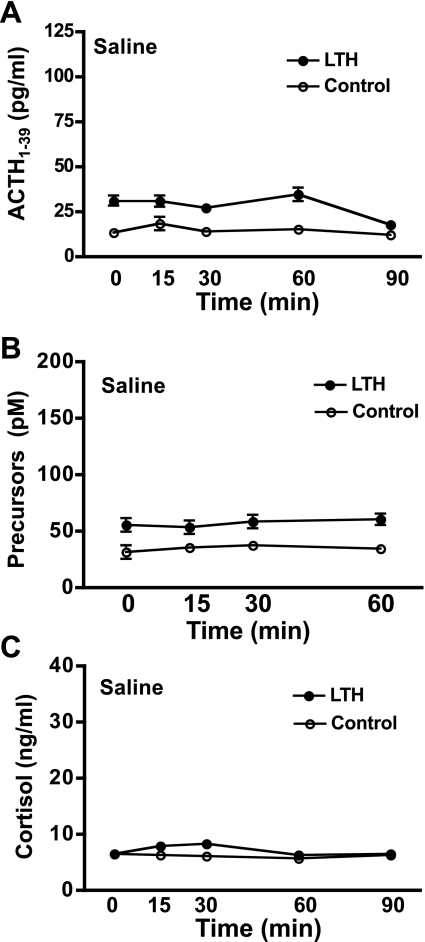
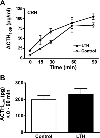
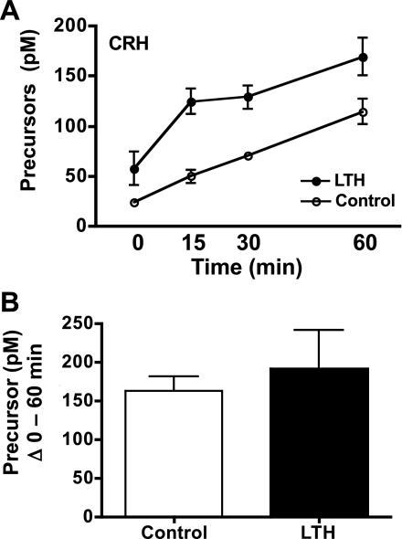
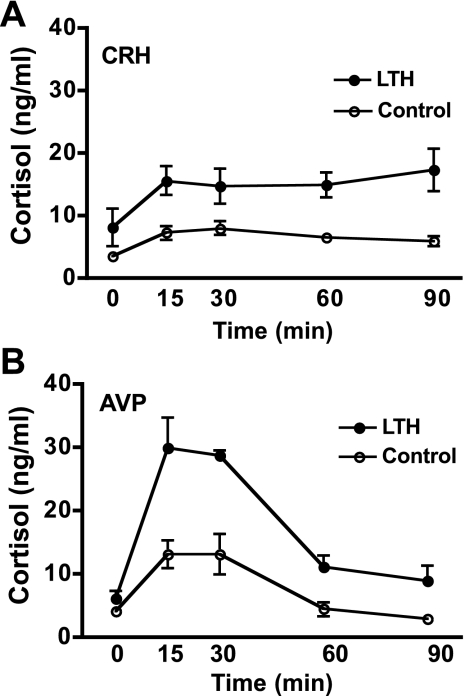
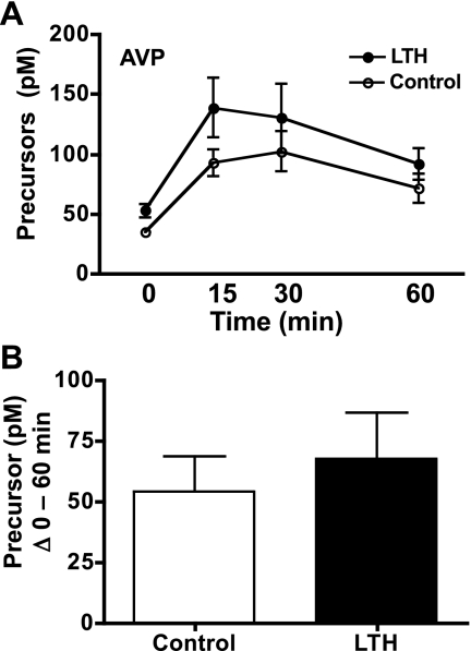
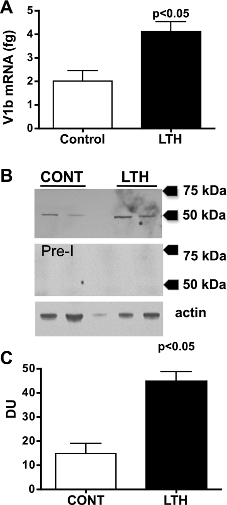
This study tested the hypothesis that long-term hypoxia (LTH) results in enhanced fetal corticotrope sensitivity to the ACTH secretagogues, corticotropin-releasing hormone (CRH), and AVP. Ewes were maintained at high altitude (3,820 m) from 40 to 130-131 days of gestation. Upon return to the laboratory, hypoxia was maintained by maternal nitrogen infusion. Vascular catheters were placed in both LTH (n = 4) and normoxic controls (n = 4). Each fetus received a 15-min infusion of either saline, 100 ng/kg of ovine CRH, or 20 ng/kg of AVP/min over 3 consecutive days in a randomized order. Fetal blood samples were collected at 0, 15, 30, 60, and 90 min after the start of infusion and analyzed for ACTH(1-39), ACTH precursors, and cortisol. Anterior pituitaries were collected from additional noninstrumented fetuses for analysis of vasopressin receptor 1b (V1b) mRNA and protein. Basal plasma concentrations of both ACTH(1-39) and ACTH precursors were higher in LTH fetuses and were not altered by saline infusion. In response to CRH, ACTH(1-39) increased in both groups and was higher in the LTH group compared with control (P < 0.05). When analyzed as sum of ACTH(1-39) released (Delta0-90 min) above basal, CRH released equal amounts of ACTH(1-39) in both groups. In LTH fetuses, AVP evoked a greater ACTH(1-39) release (P < 0.05) when analyzed as an increased sum of ACTH(1-39) (Delta0-90 min) above basal. Both CRH and AVP elicited a release of ACTH precursors with no differences observed between LTH and control. AVP and CRH elicited significant increases in cortisol, which were higher in response to AVP than CRH. V1b mRNA and protein were elevated in the anterior pituitary of LTH fetuses compared with control. LTH significantly increases pituitary sensitivity to AVP. This enhanced sensitivity may be a mechanism of our previously observed enhanced corticotrope function.


Figures

Fig. 1.
Fig. 1.
Dose-response curve of the two-site ACTH1-39 ELISA for ACTH1-39, recombinant ovine 22-kDa pro-ACTH and recombinant ovine POMC. 22 kDa of pro-ACTH exhibits ∼10% cross-reactivity, while POMC exhibits ∼1% cross-reactivity.
Fig. 2.
Fig. 2.
Plasma concentrations of ACTH1-39 (A), ACTH precursors (B), and cortisol (C) in long-term hypoxic (LTH; n = 4) and control (n = 4) fetal sheep during infusion of saline as described in methods (means ± SE). Blood samples were collected at the indicated times. As analyzed by ANOVA, basal plasma concentrations of ACTH1-39 and ACTH precursors were significantly elevated in LTH compared with control fetal sheep (P < 0.05). There were no differences in plasma cortisol between the two groups. Saline infusion did not elevate ACTH1–39, ACTH precursors, or cortisol in either group.
Fig. 3.
Fig. 3.
A: plasma concentrations of ACTH1-39 in LTH and control fetal sheep during infusion of CRH. CRH infusion was initiated immediately after the preinfusion sample (n = 4 per group; means ± SE). Concentrations of ACTH1-39 were significantly elevated (P < 0.05) in response to CRH in both groups. ACTH1-39 was significantly higher (P < 0.05) in LTH compared with control fetuses prior to and during CRH infusion. B: sum of fetal plasma ACTH1-39 for LTH and control fetuses from 0 (pre-CRH) through 90 min of CRH infusion. There were no differences in total ACTH1-39 released between groups.
Fig. 4.
Fig. 4.
A: plasma concentrations of ACTH precursors in LTH and control fetal sheep during infusion of CRH. CRH infusion was initiated immediately after the preinfusion sample (n = 4 per group; means ± SE). Concentrations of ACTH precursors were significantly elevated (P < 0.05) in response to CRH in both groups. ACTH precursors were significantly higher (P < 0.05) in LTH compared with control fetuses prior to and during CRH infusion. B: sum of fetal plasma ACTH-precursors for LTH and control fetuses from 0 (pre-CRH) through 60 min of CRH infusion. There were no differences in total ACTH precursors released between groups.
Fig. 5.
Fig. 5.
A: plasma cortisol concentrations achieved in response to CRH infusion in LTH and control fetal sheep (n = 4 per group; means ± SE). Cortisol concentrations were similar prior to CRH infusion (time 0) and significantly increased in response to CRH infusion in both groups (P < 0.05). Cortisol concentrations were significantly higher (P < 0.05) in response to CRH in LTH fetal plasma compared with control for the duration of CRH infusion. B: plasma cortisol concentrations achieved in response to AVP infusion in LTH and control fetal sheep (n = 4 per group; means ± SE). Cortisol concentrations were similar prior to AVP infusion (time 0) and significantly increased in response to AVP infusion in both groups (P < 0.05). Cortisol concentrations were significantly higher (P < 0.05) in response to AVP in LTH fetal plasma compared with control for the duration of AVP infusion.
Fig. 6.
Fig. 6.
A: plasma concentrations of ACTH1-39 in LTH and control fetal sheep during infusion of AVP. AVP infusion was initiated immediately after the preinfusion sample (n = 4 per group; means ± SE). Concentrations of ACTH1-39 were significantly elevated (P < 0.05) in response to AVP in both groups. ACTH1-39 was significantly higher (P < 0.05) in LTH compared with control fetuses prior to and at the 15, 30, and 60 min times during AVP infusion. B: sum of fetal plasma ACTH1-39 for LTH and control fetuses from 0 (pre-AVP) through 90 min of AP infusion. AVP elicited a significantly higher total ACTH1-39 release in LTH compared with control fetuses over the sampling period (P < 0.05).
Fig. 7.
Fig. 7.
A: plasma concentrations of ACTH precursors in LTH and control fetal sheep during infusion of AVP. AVP infusion was initiated immediately after the preinfusion sample (n = 4 per group; mean ± SE). Concentrations of ACTH precursors were significantly elevated (P < 0.05) in response to AVP in both groups. ACTH precursors were significantly higher (P < 0.05) in LTH compared with control fetuses prior to and during AVP infusion. B: sum of fetal plasma ACTH precursors for LTH and control fetuses from 0 (pre-AVP) through 60 min of AVP infusion. There were no differences in total ACTH precursors released between groups.
Fig. 8.
Fig. 8.
A: V1b receptor mRNA in anterior pituitaries from LTH and control fetal sheep (n = 6/group; means ± SE) as determined by quantitative RT-PCR. Messenger RNA is expressed in fg V1b mRNA per 100 ng input RNA. B: Western blot analysis for the V1b receptor for two representative samples each from control (CONT) and LTH anterior pituitaries. The middle panel is a Western blot using the preimmune serum from the same rabbit used to generate the anti-ovine V1b antibody. The lower panel is a Western blot for actin demonstrating the same samples. C: densitometry for the V1b Western (n = 4 anterior pituitaries/group). V1b protein was significantly elevated in the anterior pituitaries of LTH fetal sheep compared with control (P < 0.05).

Similar articles

Cited by

References

    1. Adachi K, Umezaki H, Kaushal KM, Ducsay CA. Long-term hypoxia alters ovine fetal endocrine and physiological responses to hypotension. Am J Physiol Regul Integr Comp Physiol 287: R209–R217, 2004. - PubMed
    1. Behan DP, De Souza EB, Lowry PJ, Potter E, Sawchenko P, Vale WW. Corticotropin-releasing factor (CRF) binding protein: a novel regulator of CRF and related peptides. Front Neuroendocrinol 16: 362–382, 1995. - PubMed
    1. Bell ME, Myers TR, Myers DA. Expression of proopiomelanocortin and prohormone convertase-1 and -2 in the late-gestation fetal sheep pituitary. Endocrinology 139: 5135–5143, 1998. - PubMed
    1. Carr GA, Jacobs RA, Young IR, Schwartz J, White A, Crosby S, Thorburn GD. Development of adrenocorticotropin-(1–39) and precursor peptide secretory responses in the fetal sheep during the last third of gestation. Endocrinology 136: 5020–5027, 1995. - PubMed
    1. Castro MI, Valego NK, Zehnder TJ, Rose JC. The ratio of plasma bioactive to immunoreactive ACTH-like activity increases with gestational age in the fetal lamb. J Dev Physiol 18: 193–201, 1992. - PubMed

Publication types

MeSH terms

LinkOut - more resources