. 2009 Jul-Aug;3(4):268-73.
doi: 10.4161/chan.3.4.9342. Epub 2009 Jul 24.

The cardiac alpha(1C) subunit can support excitation-triggered Ca2+ entry in dysgenic and dyspedic myotubes

Affiliations

The cardiac alpha(1C) subunit can support excitation-triggered Ca2+ entry in dysgenic and dyspedic myotubes

Roger A Bannister et al. Channels (Austin). 2009 Jul-Aug.

Abstract

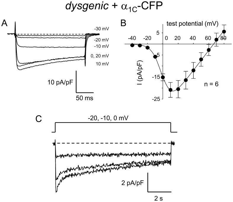
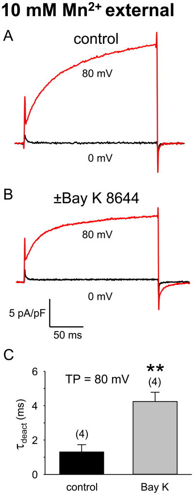
Depolarization-induced entry of divalent ions into skeletal muscle has been attributed to a process termed Excitation-Coupled Ca(2+) Entry (ECCE), which is hypothesized to require the interaction of the ryanodine receptor (RyR1), the L-type Ca(2+) channel (DHPR) and another unidentified cation channel. Thus, ECCE is absent in myotubes lacking either the DHPR (dysgenic) or RyR1 (dyspedic). Furthermore, ECCE, as measured by Mn(2+) quench of Fura-2, is reconstituted by expression of a mutant DHPR alpha(1S) subunit (SkEIIIK) thought to be impermeable to divalent cations. Previously, we showed that the bulk of depolarization-induced Ca(2+) entry could be explained by the skeletal L-type current. Accordingly, one would predict that any Ca(2+) current similar to the endogenous current would restore such entry and that this entry would not require coupling to either the DHPR or RyR1. Here, we show that expression of the cardiac alpha(1C) subunit in either dysgenic or dyspedic myotubes does result in Ca(2+) entry similar to that ascribed to ECCE. We also demonstrate that, when potentiated by strong depolarization and Bay K 8644, SkEIIIK supports entry of Mn(2+). These results strongly support the idea that the L-type channel is the major route of Ca(2+) entry in response to repetitive or prolonged depolarization of skeletal muscle.


Figures

Figure 1
Figure 1
Long, weak depolarizations in 2 mM Ca2+ elicit slowly-inactivating L-type currents in dysgenic myotubes expressing α1C-CFP. (A) A family of L-type Ca2+ currents elicited by 200-ms depolarizations from −50 mV to the indicated test potentials is shown for an α1C-CFP expressing dysgenic myotube in 2 mM external Ca2+. (B) The average peak I–V relationship is shown for dysgenic myotubes expressing α1C-CFP (●; n = 6) elicited at 0.1 Hz by test potentials ranging from −20 mV through +80 mV in 10 mV increments, following a prepulse protocol (see Materials and Methods). The smooth curve is plotted according to Eq. 1, where Gmax = 452.4 nS/nF, Vrev = 66.5 mV, V1/2 = 29.2 mV and kG = 6.42 mV. (C) Representative L-type currents from a dysgenic myotube expressing α1C-CFP elicited by 9800-ms depolarizations from −80 mV to the indicated test potentials in 2 mM external Ca2+.
Figure 2
Figure 2
ECCE-like Ca2+ entry is observed in dysgenic myotubes expressing cardiac α1C subunits. (A) Representative Ca2+ transient evoked by application of 60 mM K+ Ringer’s solution to a dysgenic myotube expressing α1C-CFP. (B) Absence of elevated K+-evoked Ca2+ transient in an untransfected dysgenic myotube. (C) Partial block by 10 μM nifedipine of the elevated K+-evoked Ca2+ transient in an α1C-CFP-expressing dysgenic myotube. (D) Complete block by 50 μM nifedipine of the K+-evoked Ca2+ transient in an α1C-CFP-expressing dysgenic myotube. (E) Average peak ΔF/F (left) and t1/2peak (right) for dysgenic myotubes expressing α1C-CFP in the absence and presence of 10 μM nifedipine. Asterisks indicate significant differences (**, p < 0.005; ***, p < 0.0005; t-test). Myotubes were exposed to 200 μM ryanodine for >1 hr at 37°C prior to experiments in order to block contribution of RyR1 and/or RyR3 to the Ca2+ transient.
Figure 3
Figure 3
ECCE-like Ca2+ entry is observed in dyspedic myotubes expressing cardiac α1C subunits. (A) Ca2+ transient in a dyspedic myotube expressing α1C-CFP evoked by 60 mM K+ Ringer’s solution. Elevated K+ failed to evoke Ca2+ transients in a non-transfected dyspedic myotube (B) or in a non-transfected dyspedic myotube exposed to 5 μM ±Bay K 8644 (C). Myotubes were exposed to 200 μM ryanodine for >1 hr at 37°C prior to experiments in order to block any potential contribution of RyR3 to the Ca2+ transient.
Figure 4
Figure 4
The SkEIIIK pore mutant allows inward Mn2+ current. Representative currents in a dysgenic myotube expressing SkEIIIK before (A) and after exposure to 5 μM ±Bay K 8644 (B). The external solution contained 10 mM Mn2+. SkEIIIK currents were elicited by depolarizations from −50 mV to either 0 or +80 mV, following a prepulse protocol (see Materials and Methods). (C) Summary of deactivation time constants of SkEIIIK Mn2+ tail currents recorded upon repolarization from 80 mV to −50 mV in the absence (n = 4) and presence (n = 4) of ±Bay K 8644. Tail currents were fit by Eq. 2. The asterisks indicate a significant difference (p < 0.006; t-test).

Similar articles

Cited by

References

    1. Cherednichenko G, Hurne AM, Fessenden JD, Lee EH, Allen PD, Beam KG, Pessah IN. Conformational activation of Ca2+ entry by depolarization of skeletal myotubes. Proc Natl Acad Sci USA. 2004;101:15793–8. - PMC - PubMed
    1. Hurne AM, O’Brien JJ, Wingrove D, Cherednichenko G, Allen PD, Beam KG, Pessah IN. Ryanodine receptor type I (RyR1) mutations C4958S and C4961S reveal excitation-coupled calcium entry (ECCE) is independent of sarcoplasmic reticulum store depletion. J Biol Chem. 2005;280:36994–7004. - PubMed
    1. Yang T, Allen PD, Pessah IN, López JR. Enhanced excitation-coupled calcium entry in myotubes is associated with expression of RyR1 malignant hyperthermia mutations. J Biol Chem. 2007;282:37471–8. - PubMed
    1. Bannister RA, Pessah IN, Beam KG. The skeletal L-type Ca2+ current is a major contributor to Excitation-Coupled Ca2+ Entry (ECCE) J Gen Physiol. 2009;133:79–91. - PMC - PubMed
    1. Cherednichenko G, Ward CW, Feng W, Carabales E, Michaelson L, Samsó M, López JR, Allen PD, Pessah IN. Enhanced Excitation-Coupled Calcium Entry (ECCE) in myotubes expressing malignant hyperthermia mutation R163C is attenuated by dantrolene. Mol Pharmacol. 2008;73:1203–12. - PMC - PubMed

Publication types

MeSH terms

LinkOut - more resources