

The neurocircuitry of fear, stress, and anxiety disorders
- PMID: 19625997
- PMCID: PMC3055419
- DOI: 10.1038/npp.2009.83
The neurocircuitry of fear, stress, and anxiety disorders
Abstract
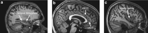
Anxiety disorders are a significant problem in the community, and recent neuroimaging research has focused on determining the brain circuits that underlie them. Research on the neurocircuitry of anxiety disorders has its roots in the study of fear circuits in animal models and the study of brain responses to emotional stimuli in healthy humans. We review this research, as well as neuroimaging studies of anxiety disorders. In general, these studies have reported relatively heightened amygdala activation in response to disorder-relevant stimuli in post-traumatic stress disorder, social phobia, and specific phobia. Activation in the insular cortex appears to be heightened in many of the anxiety disorders. Unlike other anxiety disorders, post-traumatic stress disorder is associated with diminished responsivity in the rostral anterior cingulate cortex and adjacent ventral medial prefrontal cortex. Additional research will be needed to (1) clarify the exact role of each component of the fear circuitry in the anxiety disorders, (2) determine whether functional abnormalities identified in the anxiety disorders represent acquired signs of the disorders or vulnerability factors that increase the risk of developing them, (3) link the findings of functional neuroimaging studies with those of neurochemistry studies, and (4) use functional neuroimaging to predict treatment response and assess treatment-related changes in brain function.
Figures
References
-
- Abelson JL, Khan S, Liberzon I, Young EA. HPA axis activity in patients with panic disorder: review and synthesis of four studies. Depress Anxiety. 2007;24:66–76. - PubMed
-
- Abelson JL, Liberzon I, Young EA, Khan S. Cognitive modulation of the endocrine stress response to a pharmacological challenge in normal and panic disorder subjects. Arch Gen Psychiatry. 2005;62:668–675. - PubMed
-
- Albert CM, Chae CU, Rexrode KM, Manson JE, Kawachi I. Phobic anxiety and risk of coronary heart disease and sudden cardiac death among women. Circulation. 2005;111:480–487. - PubMed
Publication types
MeSH terms
LinkOut - more resources
Full Text Sources
Other Literature Sources
Medical
