. 2009 Aug 20;460(7258):1035-9.
doi: 10.1038/nature08229. Epub 2009 Jul 22.

XIAP discriminates between type I and type II FAS-induced apoptosis

Affiliations

XIAP discriminates between type I and type II FAS-induced apoptosis

Philipp J Jost et al. Nature. .

Abstract

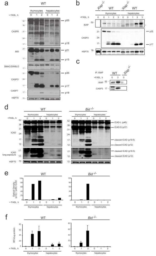
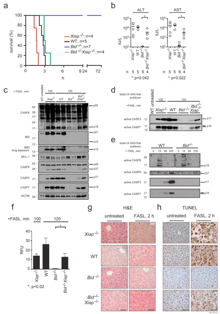
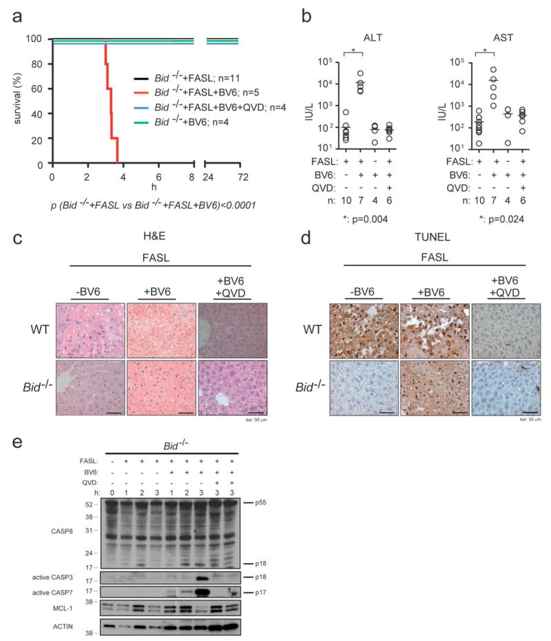
FAS (also called APO-1 and CD95) and its physiological ligand, FASL, regulate apoptosis of unwanted or dangerous cells, functioning as a guardian against autoimmunity and cancer development. Distinct cell types differ in the mechanisms by which the 'death receptor' FAS triggers their apoptosis. In type I cells, such as lymphocytes, activation of 'effector caspases' by FAS-induced activation of caspase-8 suffices for cell killing, whereas in type II cells, including hepatocytes and pancreatic beta-cells, caspase cascade amplification through caspase-8-mediated activation of the pro-apoptotic BCL-2 family member BID (BH3 interacting domain death agonist) is essential. Here we show that loss of XIAP (X-chromosome linked inhibitor of apoptosis protein) function by gene targeting or treatment with a second mitochondria-derived activator of caspases (SMAC, also called DIABLO; direct IAP-binding protein with low pI) mimetic drug in mice rendered hepatocytes and beta-cells independent of BID for FAS-induced apoptosis. These results show that XIAP is the critical discriminator between type I and type II apoptosis signalling and suggest that IAP inhibitors should be used with caution in cancer patients with underlying liver conditions.


Figures

Figure 1
Figure 1. Comparison of the levels of XIAP, caspase activation and proteolysis of caspase substrates between FASL-treated thymocytes and hepatocytes
a, Thymocytes from WT mice were treated in culture for the times indicated with 100 ng/mL FLAG-tagged FASL crosslinked with anti-FLAG antibody (2 μg/mL). Livers were harvested from WT mice that had been injected i.v. with 0.25 mg/kg FLAG-tagged FASL crosslinked with anti-FLAG antibody (2 μg/μg FASL) and sacrificed at the indicated time points. Lysates from thymocytes and livers were examined by immunoblotting for the expression and processing of caspase-8, BID, SMAC/DIABLO, caspase-3, caspase-7 and HSP70 (loading control). b, Lysates from thymocytes and hepatocytes of the indicated genotype treated as described in a were examined by immunoblotting using antibodies to XIAP, caspase-3 and HSP70 (loading control). Asterix indicates cross-reactive band. c, The binding of active caspase-3 to XIAP in liver lysates from mice of the indicated genotype treated as described in a was examined by co-immunoprecipitation with an antibody to XIAP followed by immunoblotting with antibodies to active caspase-3 or, as a loading control, with antibodies to XIAP. d, Lysates from thymocytes or hepatocytes of the indicated genotype treated as described in a were examined by immunoblotting using antibodies to the caspase substrate ICAD (lower panel exposed longer) and HSP70 (loading control). e, Relative optical density of bands (as shown in d) of cleaved ICAD (pl2) in relation to total ICAD-L (p45) is shown. f, Fluorogenic measurement of the enzymatic activity of effector caspases (DEVDase activity) in extracts from thymocytes and hepatocytes of the indicated genotype treated as described in a. Data shown are standardized per μg of protein. Three independent measurements of two individual mice per genotype and time point of treatment are shown. Error bars indicate standard error of the mean.
Figure 2
Figure 2. Loss of XIAP re-sensitises BID-deficient mice to FASL-induced fatal hepatitis
a, Long-term survival of WT, Xiap-/-, Bid-/- and Bid-/-Xiap-/- mice injected i.v. with 0.25 mg/kg FLAG-tagged FASL crosslinked with anti-FLAG antibody (2 μg/μg of FASL) is shown, p (WT vs Bid-/-)<0.0003; p (Bid-/- vs Bid-/-Xiap-/-)= 0.0005; p (WT vs Bid-/-Xiap-/-)=0.1133. b, Serum levels of ALT and AST after 120 min of FASL treatment (100 min for Xiap-/- mice) were quantified in mice of the indicated genotypes. The horizontal bars indicate the mean; n: numbers of mice analysed. P values are indicated below graphs. c, Processing of caspases -3, -7, -8, -9, BID (truncated tBID panel exposed longer), MCL-1 and β-actin (loading control) in livers of the mice treated as described in a was examined by immunoblotting. Total protein lysates were prepared from livers of two experimental animals per genotype and time point of treatment. d, Activation of caspases -3 and -7 in livers of WT, Xiap-/-, Bid-/- and Bid-/-Xiap-/- mice treated as described in a and sacrificed at 120 min (100 min for Xiap-/- mice) was examined by pulldown with the caspase inhibitor biotin-X-VAD-fmk followed by detection by immunoblotting using caspase-specific antibodies. e, Activation of caspases -3, -7, -8 and -9 in liver extracts from WT or Bid-/- mice treated as described in a and sacrificed at the indicated time points, was examined as described in d. Asterix indicates a cross-reactive band. f, Fluorogenic measurement of the enzymatic activity of effector caspases (DEVDase activity) in the livers of the mice of the indicated genotypes treated as described in a. Four independent measurements of two individual mice per genotype and time point of treatment are shown. Data represent means +/-SEM. g, Histological analysis (H&E staining) of liver sections from animals of the indicated genotypes treated as described in a sacrificed after 120 min of FASL treatment (100 min for Xiap-/- mice) and untreated control animals. h, TUNEL staining of liver sections from the animals described in f. Images are representative of a least 3 mice per genotype and time point of treatment. Scale bar = 50 μm.
Figure 3
Figure 3. Caspase inhibitors protect Bid-/-Xiap-/- hepatocytes from FASL-induced apoptosis
a, Long-term survival of WT, Xiap-/-, Bid-/- and Bid-/-Xiap-/- mice injected i.v. with 0.25 mg/kg FLAG-tagged FASL crosslinked with anti-FLAG antibody (2 μg/μg of FASL) with or without pre-treatment with the broad spectrum caspase inhibitor Q-VD-oph (20 mg/kg, i.p., 30 min prior to FASL administration) is shown. P values are indicated below individual panels. b, Levels of transaminases ALT and AST were quantified in sera of mice of the indicated genotypes after 120 min of FASL treatment (100 min for Xiap-/- mice). The horizontal bars indicate the mean; n: numbers of mice analysed. P values as indicated by asterix. c, Histological analysis (H&E staining) of liver sections from animals of the indicated genotypes treated as described in a and sacrificed after 120 min (100 min for Xiap-/- mice). d, TUNEL staining of liver sections from animals described in c. Images are representative of a least 3 mice per genotype and time point of FASL treatment. Scale bar = 50 μm. e, Processing of caspases -3, -7, -8, -9, BID, MCL-1 and β-actin (loading control) in extracts of livers from WT and Bid-/-Xiap-/- mice treated as described in a was examined by immunoblotting.
Figure 4
Figure 4. The SMAC/DIABLO mimetic drug BV6 sensitises BID-deficient mice to FASL-induced hepatocyte destruction
a, Long-term survival of Bid-/- mice injected i.v. with 0.25 mg/kg FLAG-tagged FASL crosslinked with anti-FLAG antibody (2 μg/μg of FASL) with or without co-injection of the SMAC/DIABLO mimetic drug BV6 (10 mg/kg, i.p., 30 min prior to FASL administration) and with or without pre-treatment with the caspase inhibitor QVD-oph (20 mg/kg, i.p., 30 min prior to FASL administration) is shown. P value is indicated below graph. b, Serum levels of ALT and AST were quantified after 120 min of the indicated treatment. The horizontal bars indicate the mean; n: numbers of mice analysed. P values as indicated by asterix. c, Histological analysis (H&E staining) of liver sections from WT and Bid-/- mice that had been treated as described in a and sacrificed after 180 min of FASL treatment (120 min for all images of sections from animals that were treated with BV6 alone). d, TUNEL staining of liver sections from mice described in c. Images are representative of a least 3 mice per genotype and time point of treatment. Scale bar = 50 μm. e, Processing of caspases -3, -7, -8, MCL-1 and β-actin (loading control) in liver extracts of Bid-/- mice that had been treated as described in a and sacrificed at the indicated time points were examined by immunoblotting.

Comment in

  • Shortcut to death.
    Schattenberg JM, Schuchmann M. Schattenberg JM, et al. Hepatology. 2009 Dec;50(6):2040-3. doi: 10.1002/hep.23400. Hepatology. 2009. PMID: 19937700 No abstract available.

References

    1. Nagata S. Fas ligand-induced apoptosis. Annual Review of Genetics. 1999;33:29–55. - PubMed
    1. Krammer PH. CD95’s deadly mission in the immune system. Nature. 2000;407:789–795. - PubMed
    1. Peter ME, et al. The CD95 receptor: apoptosis revisited. Cell. 2007;129:447–450. - PubMed
    1. Strasser A, Jost PJ, Nagata S. The many roles of FAS receptor signaling in the immune system. Immunity. 2009;30:180–192. - PMC - PubMed
    1. Wang K, Yin X-M, Chao DT, Milliman CL, Korsmeyer SJ. BID: a novel BH3 domain-only death agonist. Genes and Development. 1996;10:2859–2869. - PubMed

Publication types

MeSH terms