. 2009 Nov;23(11):2109-17.
doi: 10.1038/leu.2009.143. Epub 2009 Jul 23.

A robust xenotransplantation model for acute myeloid leukemia

Affiliations

A robust xenotransplantation model for acute myeloid leukemia

P V Sanchez et al. Leukemia. 2009 Nov.

Abstract

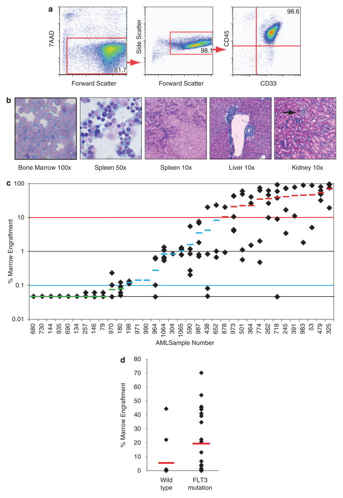
Xenotransplantation of human acute myeloid leukemia (AML) in immunocompromised animals has been critical for defining leukemic stem cells. However, existing immunodeficient strains of mice have short life spans and low levels of AML cell engraftment, hindering long-term evaluation of primary human AML biology. A recent study suggested that NOD/LtSz-scid IL2Rgammac null (NSG) mice have enhanced AML cell engraftment, but this relied on technically challenging neonatal injections. Here, we performed extensive analysis of AML engraftment in adult NSG mice using tail vein injection. Of the 35 AML samples analyzed, 66% showed bone marrow engraftment over 0.1%. Further, 37% showed high levels of engraftment (>10%), with some as high as 95%. A 2-44-fold expansion of AML cells was often seen. Secondary and tertiary recipients showed consistent engraftment, with most showing further AML cell expansion. Engraftment did not correlate with French-American-British subtype or cytogenetic abnormalities. However, samples with FLT3 mutations showed a higher probability of engraftment than FLT3 wild type. Importantly, animals developed organomegaly and a wasting illness consistent with advanced leukemia. We conclude that the NSG xenotransplantation model is a robust model for human AML cell engraftment, which will allow better characterization of AML biology and testing of new therapies.


Conflict of interest statement

Conflict of interest

Dr. Carroll has received research support from Sanofi Aventis Corporation.

Figures

Figure 1
Figure 1
Robust engraftment of primary human AML in NSG mice. (a) Representative flow cytometric analysis of bone marrow of an adult NSG mouse. 12 weeks after transplantation with 5×10⁶ human AML cells injected through tail vein 24 h after sublethal irradiation. The bone marrow is completely replaced by CD45+ CD33+ double positive cells consistent with human AML. (b) Cytospin with H & E staining of bone marrow at ×100. Cytospin with H & E staining of spleen at ×50. Spleen H & E staining at ×10 with leukemic infiltrate. Liver H & E staining at ×10 showing sinusoidal pattern of leukemic infiltrate. Kidney H & E staining at ×10, arrow shows a nest of leukemic cell infiltrate in the parenchyma of the kidney in a secondarily transplanted mouse. (c) Bone marrow engraftment of primary recipients 12–18 weeks after transplantation. 5–10×106 human AML cells from thawed pheresis samples were injected through tail vein 24 h after sublethal irradiation. The bone marrow (two tibias and two femurs) was analyzed by flow cytometry for engraftment of human AML as shown in Figure 1a. The red line indicates a high level of engraftment of>10% of murine bone marrow replaced with human AML. The blue line indicates the cutoff for engraftment. Samples with<0.1% replacement of the bone marrow with human AML were considered to be non-engrafters. Each diamond represents a single mouse. The small lines indicated in green, blue, and red indicate the average for a given sample. The samples without diamonds were analyzed as pooled samples from five individual mice. The red highlighted sample numbers indicate samples that were serially engrafted and are shown in Figure 2; 13/35 samples showed a high level of engraftment (37%), 10/35 showed a low level of engraftment (29%), and 12/35 samples showed no engraftment (34%). (d) Graphical representation of NSG engraftment (above 0.1%) based on FLT3 wild type vs FLT3 mutated samples (ITD or D835 mutation). Statistical analysis determined a 95% confidence interval of P=0.03 between engraftment of FLT3 wild type and FLT3 mutated samples.
Figure 2
Figure 2
Robust secondary and tertiary engraftment of human AML in NSG mice. (a and b) Bone marrow engraftment of secondary and tertiary recipients 12–18 weeks after serial passage; 5×10⁶ human AML cells (pooled bone marrow and spleen cells) were injected through tail vein 24 h after sublethal irradiation. Each diamond represents a single mouse. The small lines in red indicate the average for a given sample. The bone marrow is replaced by CD45+ CD33+ double positive cells consistent with human AML. There is less variability in level of engraftment between mice with serial transplant. Heavily engrafted animals show a decrease in total bone marrow engraftment because of tumor necrosis in the marrow as shown in Figure 3. (c) Fluorescence in situ hybridization probe for MLL translocation of secondarily passaged sample 391 showed evidence of the MLL fusion in 100/100 cells tested, a representative picture is shown. (d) Human AML expands in secondarily transplanted mice as analyzed by flow cytometry 12–18 weeks after transplantation. The red line indicates the average fold expansion per mouse (10×106 cells). Samples 364 had 6×10⁵ cells injected per mouse. A black circle represents the total number of CD45+ CD33+ cells from an individual mouse analysis of four bones (two tibias and two femurs) and spleen.
Figure 3
Figure 3
Leukemia-induced bone marrow fibrosis in NSG mice engrafted with human AML. (a) H & E staining of femur shows complete bone marrow replacement with leukemic infiltrate at ×10. (b) H & E staining of bone marrow shows AML tumor necrosis at ×40. (c) H & E stain shows residual fibrous debris from tumor necrosis in the bone marrow at ×40. (d) Reticulin staining shows reticulin fibrosis of the bone marrow at ×40. Collagen fibrosis of the bone marrow by trichrome staining was seen, but is not shown.
Figure 4
Figure 4
Transient co-engraftment of human cytotoxic T and AML cells in NSG mice. (a) Flow cytometric analysis of primary sample 364 after thaw shows the presence of a CD45+ CD33− population in the pheresis sample. (b) Representative flow cytometic analysis of spleen of an adult NSG mouse engrafted with sample 364, 12 weeks after transplantation with 5×10⁶ human AML cells injected through tail vein 24 h after sublethal irradiation. The spleen and bone marrow (not shown) is infiltrated by CD45+ CD33+ double positive cells consistent with human AML and a population of CD45+ CD33– cells consistent with cytotoxic T cells further characterized in (cf). (c) Spleen CD3 immunohistochemical staining at ×40 with leukemic infiltrate with scattered small and medium-sized cells staining positive for CD3 within the leukemic infiltrate. (d) Spleen CD8 immunohistochemical staining at ×40 with leukemic infiltrate with scattered small- and medium-sized cells staining positive for CD8 within the leukemic infiltrate in similar pattern to CD3 staining. (e) Spleen CD57 immunohistochemical staining at ×40 with leukemic infiltrate with scattered small- and medium-sized cells staining positive for CD57 within the leukemic infiltrate in a subset of the CD3+ cells. (f) Spleen Granzyme B immunohistochemical staining at ×40 with leukemic infiltrate with scattered small- and medium-sized cells staining positive for Granzyme B within the leukemic infiltrate in a similar pattern to CD3 staining. These findings are representative of human cytotoxic T-cell infiltration of the human leukemic infiltrate in the murine spleen. (g) Secondary passage of whole bone marrow abrogates the CD45+ CD33− population of cells in the spleen and bone marrow (not shown). (h) CD2 depletion of the primary pheresis sample before primary engraftment also abrogates expansion of the CD45+ CD33− cell population in the spleen and bone marrow (not shown).

References

    1. Ailles LE, Gerhard B, Kawagoe H, Hogge DE. Growth characteristics of acute myelogenous leukemia progenitors that initiate malignant hematopoiesis in nonobese diabetic/severe combined immunodeficient mice. Blood. 1999;94:1761–1772. - PubMed
    1. Bonnet D, Dick JE. Human acute myeloid leukemia is organized as a hierarchy that originates from a primitive hematopoietic cell. Nat Med. 1997;3:730–737. - PubMed
    1. Lapidot T, Fajerman Y, Kollet O. Immune-deficient SCID and NOD/SCID mice models as functional assays for studying normal and malignant human hematopoiesis. J Mol Med. 1997;75:664–673. - PubMed
    1. Christianson SW, Greiner DL, Hesselton RA, Leif JH, Wagar EJ, Schweitzer IB, et al. Enhanced human CD4+ T cell engraftment in beta2-microglobulin-deficient NOD-scid mice. J Immunol. 1997;158:3578–3586. - PubMed
    1. Shultz LD, Schweitzer PA, Christianson SW, Gott B, Schweitzer IB, Tennent B, et al. Multiple defects in innate and adaptive immunologic function in NOD/LtSz-scid mice. J Immunol. 1995;154:180–191. - PubMed

Publication types

Substances