. 2009 Sep;132(3):263-80.
doi: 10.1007/s00418-009-0617-x. Epub 2009 Jul 22.

Limiting transport steps and novel interactions of Connexin-43 along the secretory pathway

Affiliations

Limiting transport steps and novel interactions of Connexin-43 along the secretory pathway

Irina V Majoul et al. Histochem Cell Biol. 2009 Sep.

Abstract

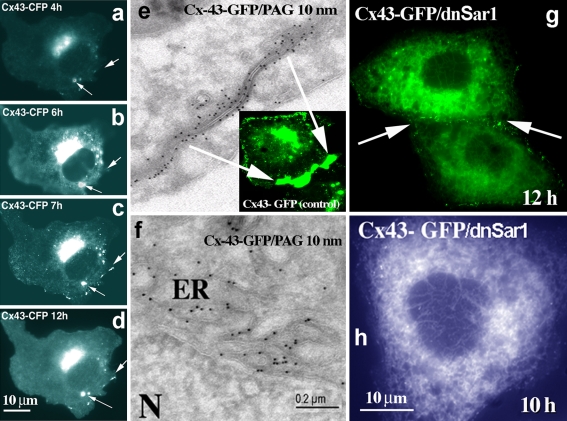
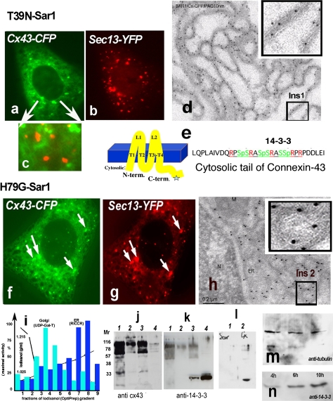
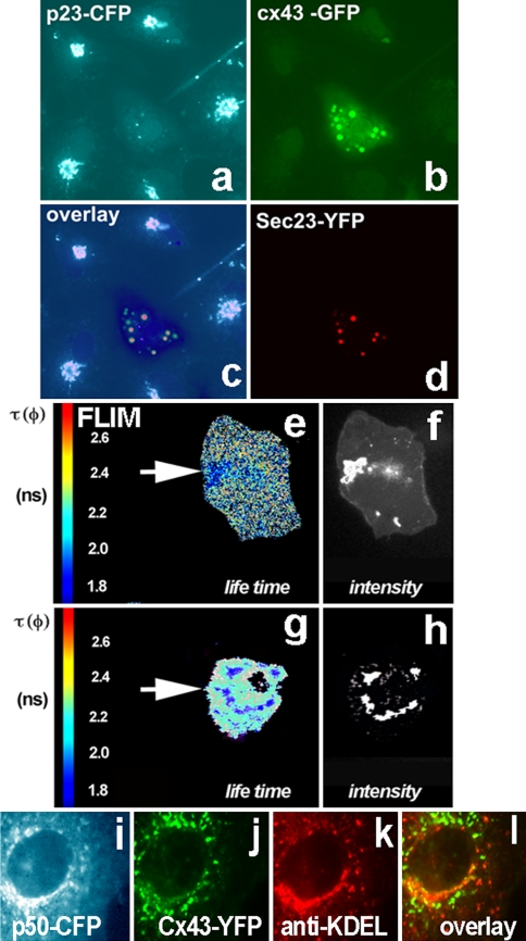
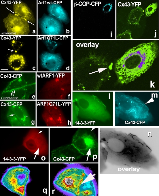
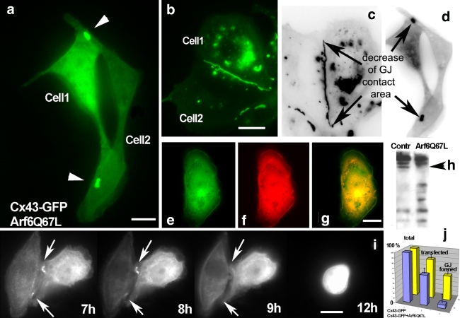
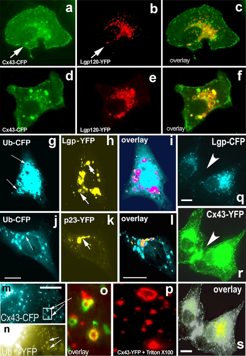
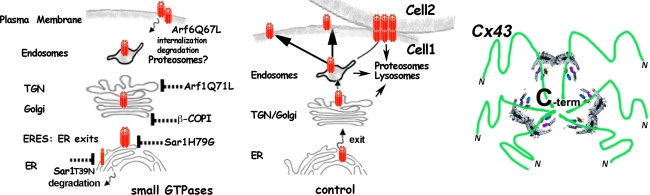
Connexins are four-transmembrane-domain proteins expressed in all vertebrates which form permeable gap junction channels that connect cells. Here, we analysed Connexin-43 (Cx43) transport to the plasma membrane and studied the effects of small GTPases acting along the secretory pathway. We show that both GTP- and GDP-restricted Sar1 prevents exit of Cx43 from the endoplasmic reticulum (ER), but only GTP-restricted Sar1 arrests Cx43 in COP II-coated ER exit sites and accumulates 14-3-3 proteins in the ER fraction. FRET-FLIM data confirm that already in ER exit sites Cx43 exists in oligomeric form, suggesting an in vivo role for 14-3-3 in Cx43 oligomerization. Exit of Cx43 from the ER can be blocked by other factors--such as expression of the beta subunit of the COP I coat or p50/dynamitin that acts on the microtubule-based dynein motor complex. GTP-restricted Arf1 blocks Cx43 in the Golgi. Lastly, we show that GTP-restricted Arf6 removes Cx43 gap junction plaques from the cell-cell interface and targets them to degradation. These data provide a molecular explanation of how small GTPases act to regulate Cx43 transport through the secretory pathway, facilitating or abolishing cell-cell communication through gap junctions.


Figures

Fig. 1
Fig. 1
Analyses of Cx43 assembly and exit from the ER. Vero cells were transfected with Cx43-CFP and analyzed for Cx43 transport at different time intervals after transfection (4–12 h). In contrast to non-contacting cells (ad), activity-dependent transport of Cx43 to the PM is facilitated by the presence of cell–cell contacts (e). Sar1H79G blocks Cx43 exit from the ER (g, f, h) and prevents formation of gap junction plaques (g). In the absence of cell–cell contacts Cx43 from the Golgi pool is delivered to distinct perinuclear dots (ad), that can be later colocalized to the lysosomal markers (see further Fig. 6). e Classical Tokuyasu ultrathin cryosections of Vero cells transfected with Cx43-GFP (12 h) were probed with anti-GFP antibodies (Invitrogen), followed by protein A-gold detection (PAG; 10-nm gold). Typical gap junction plaque between two contacting membranes is shown; f Ultrathin cryosections (Tokuyasu) of Vero cells co-transfected with Cx43-GFP and Sar1H79G reveal an ER localization. Cells were fixed 12 h after transfection and probed with anti-GFP antibodies (PAG; 10-nm gold). In contrast to control cells (e), immuno-EM analysis of cells expressing Cx43-GFP and Sar1H79G (f) reveals strong accumulation of Cx43 in the perinuclear ER region. g, h In double-transfected cells Sar1H79G blocks exit of Cx43-GFP from the ER and connexins fail to accumulate at the cell–cell interface (g, arrow); compare to control cells transfected only with Cx43-GFP (e). Scale bar 200 nm for EM and 10 μm for fluorescence microscopy images
Fig. 2
Fig. 2
ah Two distinct states of Cx43 in the ER in Vero cells expressing Sar1T39N or Sar1H79G. Only Sar1H79G accumulates Cx43 in ER exit sites (ERES) (h). This state correlates with accumulation of 14-3-3 molecules in the ER fraction (see k). a–c Cx43-GFP in cells co-transfected with Sar1T39N remains in the ER membranes, does not segregate into exit sites and does not co-localize with the COPII coat (c, overlay). d Immuno-EM analysis of ultrathin cryosections of Vero cells co-transfected with Cx43-GFP and Sar1T39N probed with anti-GFP antibodies and protein A-10 nm gold revealed that Cx43 immunoreactivity was distributed evenly along an extended ER network and not concentrated in the characteristically rounded ERES. A region of interest (Ins 1) is shown enlarged. e Cx43 transmembrane topology and acid sequence at the cytosolic C-terminus of Cx43 harboring serines in a context suitable for phosphorylation, 14-3-3 protein interaction and RPR/COPI binding motifs. fg Sar1H79G induces colocalization of Cx43 with the Sec13 subunit of the COPII coat. h Tokuyasu cryo-sections of Vero cells co-transfected with Cx43-GFP and Sar1H79G. Immunogold staining with anti-GFP antibodies show ER cisternae and clearly detectable rounded ERES structures that are decorated with Cx43-GFP. A region of interest showing Cx43 concentrated in ERES is shown (Ins 2). Co-localization between Cx43 and the COPII subunit Sec13-YFP is increased in cells co-expressing Sar1H79G (fg). i Subcellular fractionation of monolayer-grown Vero cells. ER and Golgi fractions were separated from the cytosol and PM fractions and tested for enzymatic activity: UDP-Gal-Transferase for the Golgi; Rotenon-Insensitive Cytochrome C-Reductase for the ER. Total protein concentrations in the fractions are indicated. jk Proteins from combined ER fractions 7 and 8 (i) were resolved by SDS-PAGE and immunoblotted with anti-Cx43 and Pan-14-3-3 antibodies. Lanes are as follows: 1 ER fraction of cells transfected with Sar1T39N, 2 control Golgi fraction, 3 ER fraction of cells transfected with Sar1H79G, 4 cytosol. ER fractions (7–8 of the gradient, i) usually show upper oligomeric bands of Cx43 (j) with the Cx43 antibodies; these bands are absent in the cytosol fraction (j, lane 4) (k). Note that 14-3-3 accumulation is observed only in the ER fraction of cells transfected with Sar1H79G. 14-3-3 proteins are abundantly present in cytosol, used as a positive control. l Vero cells transfected with Sar1H79G alone do not show accumulation of 14-3-3 immunoreactivity in the ER (l, 1). Accumulation of 14-3-3 proteins in the ER fraction is seen only in cells co-transfected with Cx43 and Sar1H79G (l, 2). mn Time-dependent accumulation of 14-3-3 proteins in the ER fraction of Vero cells co-transfected with Cx43 and Sar1H79G (4–10 h). The same membrane was reprobed with anti-tubulin as a loading control
Fig. 3
Fig. 3
Oligomerization of Cx43 resolved in living cells using FRET-FLIM analysis. ER-to-Golgi transport of oligomerized Cx43 requires COPII and functional dynein motor complex. a p23-CFP stably expressed in Vero cells shows a typical Golgi pattern. Such cells were used for further transfection with Cx43-GFP, Sar1H79G and Sec23-YFP (ad). The cell in the center (a) expresses all four constructs. b Cx43 is blocked in ER exit sites by Sar1H79G which also blocks p23 transport from the ER and thus prevents its Golgi accumulation (middle cell, a). Cx43 co-localizes with the COPII component Sec23-YFP (d). c Composite overlay of three channels: CFP, GFP and YFP. The fluorescence of the three channels was resolved by linear signal unmixing on the FV1000 confocal microscope. The overlay shows that in Sar1H79G expressing cells Cx43-GFP colocalizes with Sec23-YFP in ERES (resolved by EM in Fig. 2h), while the Golgi protein p23-CFP cannot be detected in the Golgi and is dispersed in the ER membrane. eh Fluorescence Life Time Microscopy (FLIM) measurements in Vero cells co-expressing the Cx43-CFP/Cx43-YFP FRET pair. e Control cells (expressing no Sar1H79G) co-transfected with the donor/acceptor pair Cx43-CFP and Cx43-YFP show accumulation of both fluorescence signals in the Golgi region. Analysis of the distribution of lifetime (τ, given in nanoseconds) in the Golgi region reveals a strong decrease of τ. Lifetime was diminished from 2.5 to 1.9 ns as a result of FRET induced by close proximity of donor/acceptor proteins, due to oligomerization and segregation of connexins. f Wide-field microscopy image confirming strong accumulation of Cx43-FP in the Golgi region. g, h In cells co-transfected with the Cx43-CFP/Cx43-YFP donor/acceptor pair and Sar1H79G, Cx43-FP fails to arrive to the Golgi and instead appears in perinuclear dots corresponding to ERES (for comparison see EM images of ERES in Fig. 2h). h The Cx43-FP labeled perinuclear structures display a similar decrease of fluorescence lifetime (2.3–1.8 ns) as those seen in the control Golgi region (g), as a result of FRET. il ER–to-Golgi traffic of Cx43 utilizes the microtubule-based, minus-end directed dynein motor complex. i Overexpression of p50-CFP, the dynamitin subunit of the dynein/dynactin complex, blocks ER-to-Golgi transport of Cx43-YFP, (j); in such cells Cx43 is unable to enter the cis/medial-Golgi detected with antibodies against KDEL-receptor (k). The overlay (l) shows absence of colocalization for Cx43 with the cis/medial-Golgi marker KDEL-receptor
Fig. 4
Fig. 4
Arf1Q71L prevents formation of gap junction plaques and blocks Cx43 exit from the Golgi. Effects of expression of β-COP and 14-3-3 expression on Cx43 transport to the Golgi. ab Cx43-CFP co-expressed with wild-type Arf1-YFP in Vero cells. Contacting cells form gap junctions (indicated by arrows). In the absence of cycloheximide Cx43-CFP can be seen in the ER, Golgi and at the PM. cd In cells expressing Arf1Q71L, Cx43-CFP fails to form gap junction plaques at the PM. e, f Cells co-expressing Cx43-CFP and wild-type Arf1-YFP and treated for 1 h with cycloheximide before imaging display Cx43 only at the PM (arrows Cx43-CFP at the cell–cell interface). g, h Cx43-CFP accumulates in the Golgi in cells co-expressing Arf1Q71L. A cell–cell contact region depleted of Cx43-CFP is indicated by arrow in g. Cells were treated for 1 h with cycloheximide. i, j Expression of β-COP-CFP results in a strong block of ER-to-Golgi transport of Cx43-YFP. k Enlarged overlay of the two channels. An ER distribution for Cx43 is observed in the cell to the right that expresses β-COP-CFP. In contrast, the cell on the left transfected only with Cx43-YFP shows a PM labeling for Cx43 at a cell–cell contact site (arrow). l, m, n 14-3-3 colocalizes with Cx43 at the edge of non-contacting PM regions in co-transfected cells. op In cells co-transfected with Cx43-CFP and 14-3-3ε-YFP, 14-3-3 reactivity never accumulates at the contacting cell–cell interface. However, the two proteins do co-localize in the Golgi (arrows). q Absence of 14-3-3ε at the cell–cell interface revealed by fluorescence intensity distribution; r In contrast, in the same cell Cx43 is clearly visible seen at the PM interface with a contacting cell (arrow)
Fig. 5
Fig. 5
Arf6-GTP diminishes formation of Cx43 gap junctions and facilitates Cx43 degradation. a Formation of gap junction plaques is strongly reduced in Vero cells co-expressing Cx43-GFP (green) and Arf6Q67L (not visualized here). The GFP signal was recorded 10 h after transfection. b Control cells expressing Cx43-GFP alone display the usual accumulation of Cx43-GFP in gap junctions at the cell–cell interface at 10 h after transfection. Inverted black–white images (c, d) more clearly reveal the relative decrease in cell surface area occupied by Cx43 gap junctions in cells expressing Arf6Q67L. Note absence of lysosomal accumulation of Cx43 in Arf6Q67L expressing cells (d) compared to control cells (c). For comparison lysosomal accumulation of Cx43 colocalized with lysosomal markers in control cells is shown in Fig. 6a–f. e, f, g In Arf6Q67L expressing cells Cx43-GFP (e) partially colocalizes with Paxillin (f) at the cell periphery (overlay in g). Note again absence of Cx43 accumulation in characteristic lysosomal structures. h Membrane fractions of control Vero cells (left) or Arf6Q67L expressing cells (right) were immunoblotted with anti-Cx43 antibodies. Strong degradation of Cx43 in the membrane fraction from ARF6Q67L expressing cells is evident. The upper (phosphorylated) forms of Cx43 are more resistant to degradation (indicated by arrowhead). i Time-dependent analyses of cells co-expressing Cx43-GFP and Arf6Q67L (time interval 7–12 h) reveal the failure of Cx43 to form gap junction plaques at cell–cell interfaces. Remaining gap junctions are degraded (compare to cells expressing Cx43-GFP alone (bc)). j Diagram of transfection rate and gap junction formation in control cells expressing Cx43-GFP alone (yellow bars) and cells co-expressing Arf6Q67L and Cx43-GFP (violet bars). Note the strong reduction in total gap junction area in the presence of Arf6Q67L
Fig. 6
Fig. 6
Cx43 transport to the cell surface and degradation in lysosomes in response to mild stress. Vero cells were co-transfected with Cx43-CFP (a) and the lysosomal marker Lgp120-YFP (b); images were taken 10 h after transfection via CFP and YFP channels, respectively. Overlay of a and b reveals only partial colocalization in the perinuclear region (c). df Cellular stress induced by exposure of transfected cells to high potassium medium (120 mM K without Ca2+) for 2 h redirects Cx43 from the PM to lysosomes. Extensive colocalization of Cx43-CFP (d) and the lysosomal marker lgp-120-YFP (e) in ring-like, typical lysosomal structures is seen in the overlay (f). g, h, i In Vero cells co-expressing Ubiquitin-CFP (g) and Lgp-120-YFP (h) these proteins colocalize in a perinuclear region and in specific rings underneath the PM (i). By EM these structures have the appearance of multivesicular bodies (data not shown). j, k, l In contrast, the Golgi protein p23-YFP (j) does not co-localize with Ubiquitin-CFP (k) upon co-expression (see overlay in l). m, n, o Cx43-CFP internalized from non-contacting PM areas (m, shown in blue) directly co-localized with Ubiquitin-YFP (n, shown in yellow) in ring-like lysosomal structures (displayed enlarged, as red/green overlay, in o). Remarkably, similar types of structures can be observed after brief treatment of cells harboring Cx43-GFP plaques with Triton X-100 (p), known to destroy all but detergent-resistant structures. q, r, s Lysosomes (Lgp120-CFP, q) are not present underneath freshly formed Cx43 plaques (Cx43-YFP, r), see arrow pointing to a Cx43-YFP-positive cell–cell contact region
Fig. 7
Fig. 7
Schematic involvement of small GTPases in Cx43 transport. Left panel Distribution and effects of three small GTPases that act in the biosynthetic pathway, Sar1, Arf1, and Arf6, on the transport of Cx43. Differential blocks induced by GDP- and GTP-restricted Sar1 on ER-to-Golgi transport of Cx43: Sar1T39N prevents assembly into ER exit sites—left, Sar1H79G blocks Cx43 in ER exit sites—right (monomeric and oligomeric forms of Cx43 are depicted in red). Cx43 exit from the Golgi is blocked by ARF1Q71L. Recycling and removal of Cx43 at the PM is Arf6-dependent. This step may be important for regulation of Cx43 at the PM in response to extracellular signals, migration, or initiation of transformation. Middle panel ER-Golgi-PM transport of connexins and formation of gap junctions at the cell–cell interface upon contact. Right panel Possible role of 14-3-3 proteins in Cx43 oligomerization/segregation into ERES

Similar articles

Cited by

References

    1. {'text': '', 'ref_index': 1, 'ids': [{'type': 'DOI', 'value': '10.1083/jcb.131.4.875', 'is_inner': False, 'url': 'https://doi.org/10.1083/jcb.131.4.875'}, {'type': 'PMC', 'value': 'PMC2200014', 'is_inner': False, 'url': 'https://pmc.ncbi.nlm.nih.gov/articles/PMC2200014/'}, {'type': 'PubMed', 'value': '7490291', 'is_inner': True, 'url': 'https://pubmed.ncbi.nlm.nih.gov/7490291/'}]}
    2. Aridor M, Bannykh SI, Rowe T, Balch WE (1995) Sequential coupling between COPII and COPI vesicle coats in endoplasmic reticulum to Golgi transport. J Cell Biol 131:875–893 - PMC - PubMed
    1. {'text': '', 'ref_index': 1, 'ids': [{'type': 'DOI', 'value': '10.1016/S0306-4522(01)00492-4', 'is_inner': False, 'url': 'https://doi.org/10.1016/s0306-4522(01)00492-4'}, {'type': 'PubMed', 'value': '11784696', 'is_inner': True, 'url': 'https://pubmed.ncbi.nlm.nih.gov/11784696/'}]}
    2. Baxter HC, Liu WL, Forster JL, Aitken A, Fraser JR (2002) Immunolocalisation of 14-3-3 isoforms in normal and scrapie-infected murine brain. Neuroscience 109:5–14 - PubMed
    1. {'text': '', 'ref_index': 1, 'ids': [{'type': 'DOI', 'value': '10.1016/S0092-8674(03)01079-1', 'is_inner': False, 'url': 'https://doi.org/10.1016/s0092-8674(03)01079-1'}, {'type': 'PubMed', 'value': '14744428', 'is_inner': True, 'url': 'https://pubmed.ncbi.nlm.nih.gov/14744428/'}]}
    2. Bonifacino JS, Glick BS (2004) The mechanisms of vesicle budding and fusion. Cell 116:153–166 - PubMed
    1. {'text': '', 'ref_index': 1, 'ids': [{'type': 'PubMed', 'value': '4473832', 'is_inner': True, 'url': 'https://pubmed.ncbi.nlm.nih.gov/4473832/'}]}
    2. Bozhkova VP, Kvavilashvili IS, Rott NN, Chailakhyan LM (1974) Measurement of electrical coupling between cells of axolotl embryos during cleavage divisions. Sov J Dev Biol 4:480–482 - PubMed
    1. {'text': '', 'ref_index': 1, 'ids': [{'type': 'DOI', 'value': '10.1083/jcb.139.2.469', 'is_inner': False, 'url': 'https://doi.org/10.1083/jcb.139.2.469'}, {'type': 'PMC', 'value': 'PMC2139801', 'is_inner': False, 'url': 'https://pmc.ncbi.nlm.nih.gov/articles/PMC2139801/'}, {'type': 'PubMed', 'value': '9334349', 'is_inner': True, 'url': 'https://pubmed.ncbi.nlm.nih.gov/9334349/'}]}
    2. Burkhardt JK, Echeverri CJ, Nilsson T, Vallee RB (1997) Overexpression of the dynamitin (p50) subunit of the dynactin complex disrupts dynein-dependent maintenance of membrane organelle distribution. J Cell Biol 139:469–484 - PMC - PubMed

Publication types