. 2010 Mar 15;126(6):1327-38.
doi: 10.1002/ijc.24767.

Rlip76 transports sunitinib and sorafenib and mediates drug resistance in kidney cancer

Affiliations

Rlip76 transports sunitinib and sorafenib and mediates drug resistance in kidney cancer

Sharad S Singhal et al. Int J Cancer. .

Abstract

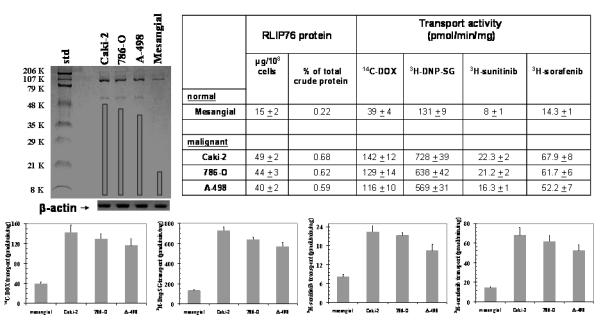
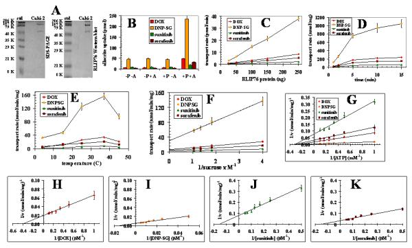
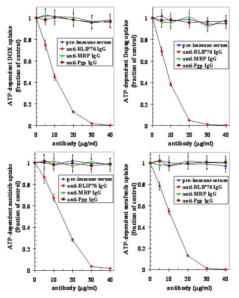
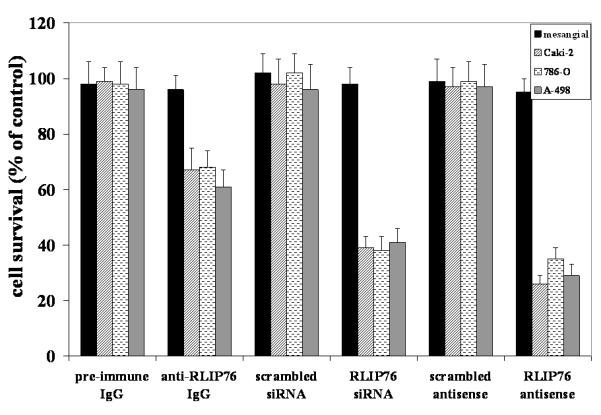
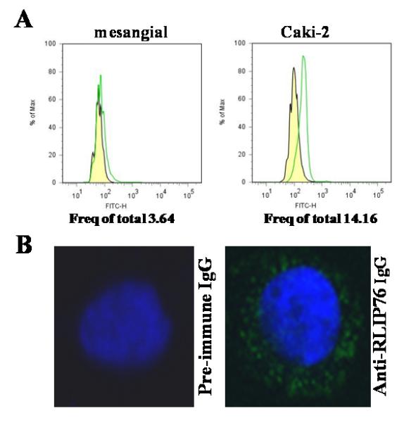
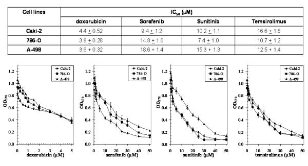
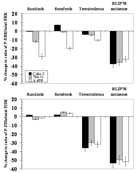
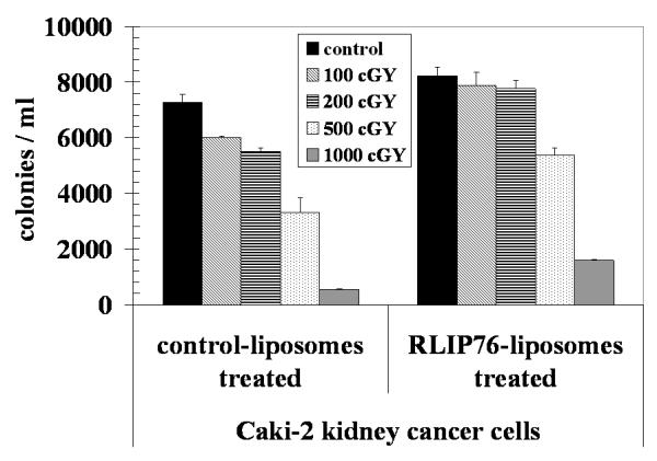
RLIP76 is a stress-responsive membrane protein implicated in the regulation of multiple cellular signaling pathways. It represents the predominant glutathione-conjugate (GS-E) transporter in cells. We have shown that RLIP76 plays a crucial role in defending cancer cells from radiation and chemotherapeutic toxin-mediated apoptosis, and that its inhibition by antibodies or depletion by siRNA or antisense causes apoptosis in a number of cancer cell types. We demonstrated for the first time that the striking anti-neoplastic effects with no evident toxicity in terms of either weight loss or metabolic effects are also demonstrable for the antibody, antisense and siRNA in a renal cell xenografts model of Caki-2 cells (Singhal et al., Cancer Res., 2009, 69: 4244). Present studies were performed to determine if RLIP76 targeting is more broadly applicable in other kidney cancer cell lines, to compare the signaling effects of RLIP76 antisense with kinase inhibitors used in treatment of renal cell carcinoma, and to determine whether kinase inhibitors were substrates for transport by RLIP76. Results of these studies show that sorafenib as well as sunitinib are substrates for transport by RLIP76 thus are competitive inhibitors of GS-E transport. Furthermore, kinase inhibition in the ERK as well as PI3K pathways by RLIP76 depletion is more profound and consistent and is more widely apparent in a number of renal carcinoma cell lines. These studies offer strong support for our overall hypothesis that RLIP76 is an overarching anti-apoptosis mechanism that, if inhibited, can be more broadly effective in the treatment of renal cell carcinoma.


Figures

Figure 1
Figure 1. Comparison of RLIP76 levels and transport activity in kidney malignant versus non-malignant cells
Aliquots of crude membrane fractions of malignant cells (Caki-2, 786-O and A-498) and non-malignant cells (mesangial), containing 200 μg protein were used for SDS-PAGE and Western-blotting against anti-RLIP76 IgG. Results were quantified by scanning densitometry. β-actin was used as an internal control. Cell lines were cultured in respective medium and homogenate was prepared from 100 × 106 cells. RLIP76 was purified from total crude membrane fraction using DNP-SG affinity, and quantified by ELISA. For transport studies, in-side-out vesicles (IOVs) were prepared from 20 × 106 cells. Transport-activity was calculated from measurements of uptake of 14C-DOX (sp. activity, 8.7 × 10⁴ cpm/nmol), 3H-DNP-SG (sp. activity, 3.6 × 10³ cpm/nmol), 3H-sunitinib (sp. activity, 4.5 × 10⁴ cpm/nmol) and 3H-sorafenib (sp. activity, 3.9 × 10⁴ cpm/nmol), into the IOVs (20 μg/ 30 μl reaction mixture) in the absence or presence of 4 mM ATP. Each transport experiment was done in triplicates in three separate experiments (n = 9). Please note the differences in scales between the panels.
Figure 2
Figure 2. Transport of 14C-DOX, 3H-DNPSG, 3H-sunitinib and 3H-sorafenib by reconstituted RLIP76 purified from Caki-2 cells
DNP-SG affinity purified RLIP76 from Caki-2 kidney cancer cells was applied to SDS-PAGE and subjected to Western blot analyses against anti-RLIP76 IgG as a primary antibody. SDS-PAGE was stained with Coomassie Brilliant Blue-R250 and Western blots were developed using horseradish peroxidase-conjugated goat-anti-rabbit-IgG as secondary antibody (panel A). Purified RLIP76 was reconstituted into artificial liposomes and uptake of radio-labeled 14C-DOX, 3H-DNPSG, 3H-sunitinib and 3H-sorafenib by control- or RLIP76-proteoliposomes were measured in the presence or absence of ATP. Vesicles equivalent to 250 ng of purified RLIP76 were used per 30 μl filtered reaction mixture in all panels except C. Total radioactivity retained by 0.45 μm filters in 96-well plates was determined after solubilizing filters in scintillation fluid, converted to pmol using the specific activity of 14C-DOX (87.4 cpm/pmol), 3H-DNPSG (3.7 cpm/pmol), 3H-sunitinib (43.5 cpm/pmol) and 3H-sorafenib (39.2 cpm / pmol), Each point represents an average and SD calculated from 12 measurements [control (−P) or RLIP76-proteoliposomes (+P), without (−A) or with ATP (+), in triplicates]. Uptake in RLIP76-proteoliposomes with ATP is significantly greater than other groups (p<0.005) (panel B). All transport studies were carried out using 250 ng protein/assay except when protein was varied (panel C). Incubation time was 5 min except for time dependence studies (panel D). Temperature was 37 °C except for temperature dependence studies (panel E). External sucrose concentration was 40 mM, except for studies of osmolar dependence (panel F). ATP was 4 mM except in ATP-dependence studies (panel G). DOX was 3.6 μM except in DOX-dependence studies (panel H). DNP-SG was 100 μM except in DNPSG-dependence studies (panel I). Sunitinib was 10 μM except in sunitinib-dependence studies (panel J). Sorafenib was 10 μM except in sorafenib-dependence studies (panel K). The proteoliposomes prepared from RLIP76 purified from Caki-2 cells. Mean values ± SD for three experiments in triplicates are shown.
Figure 3
Figure 3. Dose dependent inhibition of 14C-DOX, 3H-DnpSG, 3H-sunitinib and 3H-sorafenib transport in purified RLIP76 reconstituted vesicles from Caki-2 cells by anti-RLIP76, anti-MRP and anti-Pgp IgG
Proteoliposomes were incubated in the presence of varying concentration of antibodies (5-40 μg/ml transport reaction) for 30 min followed by determination of 14C-DOX, 3H-DnpSG, 3H-sunitinib and 3H-sorafenib uptake by proteoliposomes in the absence or presence of ATP as described in Methods section. Results presented are mean and SD from three experiments in triplicates. (◆), pre-immune serum; (●), anti-RLIP76 IgG; (▲), anti-MRP1 IgG and (■), anti-Pgp IgG.
Figure 4
Figure 4. Selective toxicity of anti-RLIP76 IgG, RLIP76-siRNA and RLIP76-antisense towards kidney cancer cells
Effect of anti-RLIP76 IgG (40 μg/ml final concentration) on cell survival was determined by MTT assay. Depletion of RLIP76 expression by RLIP76-siRNA (20 μg/ml final conc.) and RLIP76-antisense (10 μg/ml final conc.) was done, using Transmessenger Transfection Reagent kit (Qiagen), and Maxfect Transfection Reagent (Molecula, Inc.), according to the manufacturer’s instructions, respectively. Cell-survival was measured by MTT cytotoxicity assay 24 h after treatment. Values are presented as mean ± SD from two separate determinations with eight replicates each (n = 16).
Figure 5
Figure 5. Cell surface antigen analysis
Flow-cytometric analyses were carried out on cell suspensions (106 cells/ml) obtained by incubating monolayer cell cultures with Trypsin-EDTA. For the determination of cell-surface RLIP76, the mesangial (normal) as well as Caki-2 kidney cancer cells were incubated for 20 min at 4 °C with anti-RLIP76 IgG as a primary antibody. After washing with phosphate buffered saline containing NaN3, and 1% FBS, cells were incubated for 20 min at 4 °C with a FITC-conjugated goat-anti-rabbit as secondary antibody, followed by washing and than cells were immediately analyzed using BD LSR11 flow-cytometer systems using Flow Jo 7.2.2 analysis software. Yellow: pre-immune IgG + secondary antibody; Green: anti-RLIP76 IgG + secondary antibody (panel A). For cell-surface localization of RLIP76 in Caki-2 cells, cells were grown on glass cover-slips and live, unfixed cells were subjected to immuno-histochemistry using pre-immune and anti-RLIP76 IgG as the primary antibody and FITC-conjugated goat anti-rabbit IgG as the secondary antibody. DAPI was used as a nuclear counter-stain (blue-fluorescence). Slides were analyzed using a Lecia TCS SP5 confocal microscope (panel B).
Figure 6
Figure 6. DOX, Sorafenib, Sunitinib and Temsirolimus IC50 values in renal carcinoma
Drug-sensitivity assays were performed using MTT to determine IC50 values. The values are presented as mean ± SD from three separate determinations with eight replicates each (n = 24). Please note the differences in scales between the figures.
Figure 7
Figure 7. Percent change in phosphorylated ERK and PI3K caused by sorafenib, sunitinib, temsirolimus and RLIP76-antisense
Kidney cancer cells were seeded in 96 well plate (~50, 000 cells per well) and treated with 20 μM sunitinib, sorafenib and temsirolimus, and RLIP76-antisense (10 μg/ml) for 24 h and were fixed with 4% formaldehyde in PBS for 20 minutes at room temperature followed by washing with wash buffer. The vendor-provided method was used to determine total and phosphorylated proteins using phospho-specific antibodies (Active Motif). The results presented represent the percent change in phosphorylated protein compared with corresponding untreated cells.
Figure 8
Figure 8. Radiation protections by RLIP76-liposomal delivery
2.5 × 10³ Caki-2 cells were treated with control and RLIP76-liposomes (50 μg/ml final conc.) for 24 h prior to radiation at 100, 200, 500 and 1000 cGY (6 MeV photons, 1.25 min) at the Texas Cancer Center, Arlington, TX. After 7 days, cells were stained with methylene blue and the colonies were counted using an Innotech Alpha Imager HP. The results presented are the mean and s.d. from three separate experiments.

Similar articles

Cited by

References

    1. Yao M, Yoshida M, Kishida T, Nakaigawa N, Baba M, Kobayashi K, Miura T, Moriyama M, Nagashima Y, Nakatani Y, Kubota Y, Kondo K. VHL tumor suppressor gene alterations associated with good prognosis in sporadic clear-cell renal carcinoma. J Natl Cancer Inst. 2002;94:1569–75. - PubMed
    1. Ohh M, Park CW, Ivan M, Hoffman MA, Kim TY, Huang LE, Pavletich N, Chau V, Kaelin WG. Ubiquitination of hypoxia-inducible factor requires direct binding to the beta-domain of the von Hippel-Lindau protein. Nat Cell Biol. 2000;2:423–7. - PubMed
    1. Hudes G, Carducci M, Tomczak P, Dutcher J, Figlin R, Kapoor A, Staroslawska E, Sosman J, McDermott D, Bodrogi I, Kovacevic Z, Lesovoy V, et al. Temsirolimus, interferon alfa, or both for advanced renal-cell carcinoma. N Engl J Med. 2007;356:2271–81. - PubMed
    1. Thomas GV, Tran C, Mellinghoff IK, Welsbie DS, Chan E, Fueger B, Czernin J, Sawyers CL. Hypoxia-inducible factor determines sensitivity to inhibitors of mTOR in kidney cancer. Nat Med. 2006;12:122–7. - PubMed
    1. Mendel DB, Laird AD, Xin X, Louie SG, Christensen JG, Li G, Schreck RE, Abrams TJ, Ngai TJ, Lee LB, Murray LJ, Carver J, et al. In vivo antitumor activity of SU11248, a novel tyrosine kinase inhibitor targeting vascular endothelial growth factor and platelet-derived growth factor receptors: determination of a pharmacokinetic/pharmacodynamic relationship. Clin Cancer Res. 2003;9:327–37. - PubMed

Publication types

MeSH terms