. 2009 Sep 14;423(1):79-89.
doi: 10.1042/BJ20090993.

Fibulin-4 regulates expression of the tropoelastin gene and consequent elastic-fibre formation by human fibroblasts

Affiliations

Fibulin-4 regulates expression of the tropoelastin gene and consequent elastic-fibre formation by human fibroblasts

Qiuyun Chen et al. Biochem J. .

Abstract

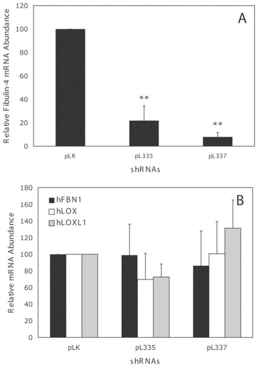
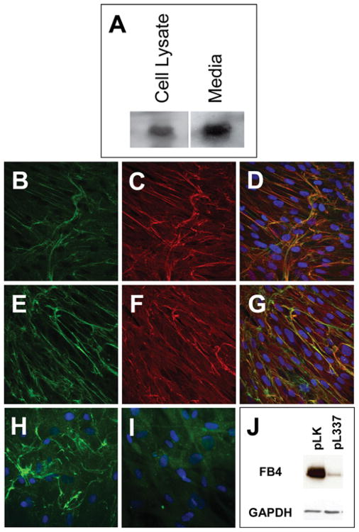
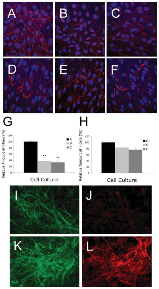
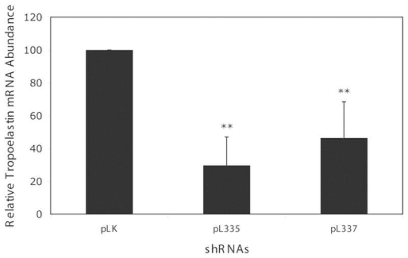
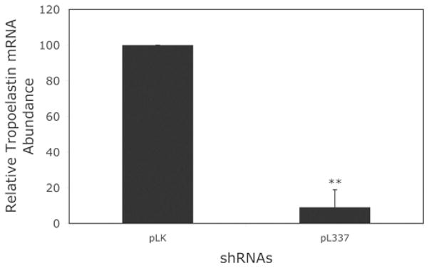
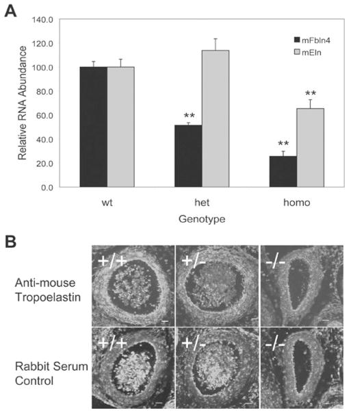
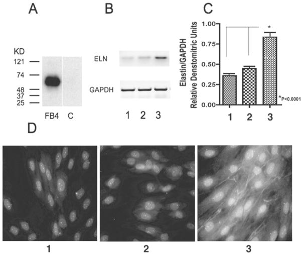
Elastic fibres are essential for normal physiology in numerous tissues, including arteries, lungs and skin. Fibulin-4 is an elastic-fibre-associated glycoprotein that is indispensable for elastic-fibre formation in mice. However, the mechanism by which fibulin-4 executes this function remains to be determined. Here, we established an in vitro functional assay system in which fibulin-4 was knocked down in human foreskin fibroblasts using siRNA (small interfering RNA) technology. With two different siRNAs, substantial knockdown of fibulin-4 was achieved, and this suppression was associated with impaired elastic-fibre formation by the fibroblasts. Real-time reverse transcription-PCR analysis showed that knockdown of fibulin-4 expression was accompanied by reduced expression of tropoelastin mRNA. Further analysis showed that this decrease was caused by transcriptional down-regulation of tropoelastin. This effect was selective, since the mRNA level of other elastic-fibre-associated proteins, including fibrillin-1, lysyl oxidase and lysyl oxidase-like-1, was not affected. Moreover, addition of conditioned medium from cultures of CHO (Chinese-hamster ovary) cells overexpressing fibulin-4 stimulated tropoelastin expression and elastic-fibre formation in cultures of Williams-Beuren-syndrome fibroblasts. Knocking down or knocking out fibulin-4 in mice led to a decrease in tropoelastin expression in the aorta. These results indicate that fibulin-4, considered as a structural protein, may also participate in regulating elastic-fibre formation in human cells through an unanticipated mechanism, namely the regulation of tropoelastin expression.


Figures

Figure 1
Figure 1. Partial amino acid sequence of the N-terminal His6–c-myc-tagged human fibulin-4 expressed by plasmid pQC0701
The expressed protein contains an N-terminal Igκ-chain signal peptide (underlined sequence). The predicted signal peptide cleavage site is indicated by a filled triangle. The N-terminus of the cleaved polypeptide has a His6 tag and a c-myc epitope tag (italicized and bold). The predicted mature form of human fibulin-4 polypeptide was placed C-terminal to the myc epitope in parenthesis. Only eight amino acid residues from both the N-terminus and the C-terminus of human fibulin-4 are shown here.
Figure 2
Figure 2. Relative mRNA abundance in fibroblasts infected with control pLK viruses or fibulin-4 shRNA viruses pL335 and pL337
Infecting HFFs with pL335 or pL337 viruses leads to a significant decrease in fibulin-4 mRNA (A), with no statistically significant change in the mRNA level (B) of fibrillin-1 (hFBN1), lysyl oxidase (hLOX), and lysyl oxidase-like-1 (hLOXL1). Results are means ± S.D. for six to eight samples. **P < 0.05 compared with the control pLK.
Figure 3
Figure 3. Characterization of fibulin-4 produced in HFF cells
(A) Western-blot analysis of fibulin-4 in HFF cell lysate and cultured medium. (BG) Confocal images of passage-10 HFF cultures immunostained with antibodies against human fibulin-4 (B, E), human elastin (C) and human fibrillin-1 (F). Co-localization of fibulin-4 with elastin and fibrillin-1 are shown in the merged images, (D) and (G) respectively. (H, I) Fluorescent images of HFF cultures infected with pLK virus (H) or pL337 virus (I) and immunostained with fibulin-4 antibody. (J) Western-blot analysis of fibulin-4 (FB4) in HFF cells infected with the control pLK lentivirus or the fibulin-4 shRNA-producing lentivirus pL337. Note that pL337 infection led to the abrogation of fibulin-4 staining in the ECM (I) and a significant decrease in the amount of fibulin-4 in the HFF cells (J).
Figure 4
Figure 4. Representative images of HFFs immunostained with antibodies against elastin (A, B and C) or fibrillin-1 (D, E and F) and HPCE cells immunostained with antibodies against fibulin-4 (I, K), elastin (J) or fibrillin-1 (L)
HFF cells were seeded on to glass coverslips in a 12-well plate with 1.8 × 10⁵ cells/well and were allowed to grow for 3 days before immunostaining. Fibroblasts infected with pLK control viruses (A) produced extensive elastic fibres, whereas fibroblasts infected with fibulin-4 shRNA viruses pL335 (B) and pL337(C) produced significantly fewer elastic fibres. No statistically significant difference in the production of fibrillin-1 microfibrils was present among fibroblast cultures infected with pLK (D), pL335 (E) and pL337 (F). (G) Quantification of elastic fibres taken from at least four fields of multiple cultures represented in (A), (B) and (C). (H) Quantification of microfibrils taken from four fields of multiple cultures represented in (D), (E) and (F). **P < 0.05 compared with the respective control pLK. (IL) HPCE cells were seeded on to glass coverslips in a 12-well plate with 4 × 10⁵ cells/well and were allowed to grow for 7 days before immunostaining. The cultures produced significant amounts of fibulin-4 (I, K) and fibrillin-1 (L). However, no elastic fibres were detectable (J).
Figure 5
Figure 5. Relative tropoelastin mRNA abundance in fibroblasts infected with control pLK viruses or fibulin-4 shRNA viruses pL335 and pL337
Infecting HFFs with either pL335 or pL337 viruses led to a significant decrease in tropoelastin mRNA. Results are means±S.D. for six to eight samples. **P < 0.05 compared with the control pLK. The results are based on eight samples from three experiments. Some variability in the magnitude of tropoelastin mRNA expression was observed and later traced to the batches of FBS used.
Figure 6
Figure 6. Relative tropoelastin mRNA abundance in rASMCs infected with control pLK viruses or fibulin-4 shRNA virus pL337
Infecting rASMCs with pL337 virus leads to a significant decrease in tropoelastin mRNA, as analysed by real-time RT–PCR. Results are means ± S.D. for eight samples. **P < 0.05 compared with the control pLK.
Figure 7
Figure 7. Decreased tropoelastin expression in the aorta of fibulin-4-knockdown and fibulin-4-knockout mice
(A) Real-time RT–PCR analysis of fibulin-4 (mFbln4) and tropoelastin (mEln) mRNA in the aorta of 10-day-old fibulin-4-knock-down mice. Results are means ± S.E.M. for nine to eleven samples. **P < 0.05 compared with the wild-type (wt) littermates. het, heterozygous; homo, homozygous. (B) Immunostaining images of E13.5 aorta isolated from fibulin-4-knockout mice. Top panel, stained with tropoelastin antibodies; Bottom panel, stained with control rabbit serum. The experiment was repeated at least three times with consistent results.
Figure 8
Figure 8. Changes over time of relative tropoelastin mRNA abundance in fibroblasts infected with control pLK virus or fibulin-4 shRNA viruses pL335 and pL337
The rate of tropoelastin mRNA degradation in pL335- or pL337-infected fibroblasts was not significantly different from that in pLK-infected cells as determined by Student’s t test. Results are means ± S.D. for five to eight samples.
Figure 9
Figure 9. Relative amount of tropoelastin pre-mRNA in fibroblasts infected with control pLK viruses or fibulin-4 shRNA viruses pL335 and pL337
Fibroblasts infected with pL335 or pL337 produced a significant decreased amount of tropoelastin pre-mRNA compared with that in the pLK-infected cells, indicating that the transcriptional activity of tropoelastin significantly decreased in the pL335- or pL337-infected cells as compared with that in the pLK-infected cells. Results are means ± S.D. for four or five samples. **P < 0.05 compared with the control pLK.
Figure 10
Figure 10. Effects of fibulin-4 on the elastogenesis of WBS fibroblasts
(A) Western-blot analysis of fibulin-4 in CHO-cell conditioned medium. Fibulin-4 was detected only in the conditioned medium (lane FB4) of fibulin-4-overexpressing cells and not in the conditioned medium (lane C) of control cells. (B, C) Expression of tropoelastin and GAPDH mRNAs in the WBS fibroblast cultures in DMEM with 2 % FBS (lane 1), in conditioned medium from control CHO cells mixed with equal volume of DMEM with 2 % FBS (lane 2) or in conditioned medium from CHO cells overexpressing fibulin-4 cells mixed with equal volume of DMEM with 2 % FBS (lane 3). Fibroblasts exposed to fibulin-4-overexpressing conditioned medium demonstrated a significantly higher level of tropoelastin mRNA. The experiment was repeated three times. **P < 0.0001 compared with lane 1 or lane 2. (D) WBS fibroblasts maintained for 5 days either in DMEM containing 2 % FBS or in the medium consisting of equal volumes of DMEM and conditioned medium from control CHO cell cultures did not form elastic fibres (D, panel 1) and (D, panel 2). In contrast, the parallel cultures of WBS fibroblasts maintained in medium consisting of equal volumes of DMEM and conditioned medium from cultures of CHO cells overexpressing fibulin-4 (final concn. of fibulin-4: 10 ng/ml) demonstrated a significant formation of immunodetectable elastic fibres (D, panel 3).

Similar articles

Cited by

References

    1. Mecham R, Davis E. Elastic fibre structure and assembly. In: Yurchenco P, Birk D, Mecham R, editors. Extracellular Matrix Assembly and Structure. Academic Press; San Diego: 1994. pp. 281–314.
    1. Vrhovski B, Weiss AS. Biochemistry of tropoelastin. Eur J Biochem. 1998;258:1–18. - PubMed
    1. Kielty CM, Sherratt MJ, Shuttleworth CA. Elastic fibres. J Cell Sci. 2002;115:2817–2828. - PubMed
    1. Hinek A, Pshezhetsky AV, von Itzstein M, Starcher B. Lysosomal sialidase (neuraminidase-1) is targeted to the cell surface in a multiprotein complex that facilitates elastic fibre assembly. J Biol Chem. 2006;281:3698–3710. - PubMed
    1. Hinek A. Nature and the multiple functions of the 67-kD elastin-/laminin binding protein. Cell Adhes Commun. 1994;2:185–193. - PubMed

Publication types

MeSH terms