

Tensile stress patterns predicted in the articular disc of the human temporomandibular joint
- PMID: 19627392
- PMCID: PMC2766058
- DOI: 10.1111/j.1469-7580.2009.01127.x
Tensile stress patterns predicted in the articular disc of the human temporomandibular joint
Abstract
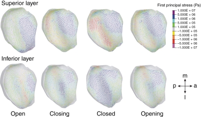
The direction of the first principal stress in the articular disc of the temporomandibular joint was predicted with a biomechanical model of the human masticatory system. The results were compared with the orientation of its collagen fibers. Furthermore, the effect of an active pull of the superior lateral pterygoid muscle, which is directly attached to the articular disc, was studied. It was hypothesized that the markedly antero-posterior direction of the collagen fibers would be reflected in the direction of the tensile stresses in the disc and that active pull of the superior lateral pterygoid muscle would augment these tensions. It was found that the tensile patterns were extremely dependent on the stage of movement and on the mandibular position. They differed between the superior and inferior layers of the disc. The hypothesis could only be confirmed for the anterior and middle portions of the disc. The predicted tensile principal stresses in the posterior part of the disc alternated between antero-posterior and medio-lateral directions.
Figures


Similar articles
-
Three-dimensional finite element analysis of the cartilaginous structures in the human temporomandibular joint.J Dent Res. 2001 Oct;80(10):1913-8. doi: 10.1177/00220345010800101001. J Dent Res. 2001. PMID: 11706951
-
Finite element analysis of stresses in the maxillary and mandibular dental arches and TMJ articular discs during clenching into maximum intercuspation, anterior and unilateral posterior occlusion.Stomatologija. 2007;9(4):121-8. Stomatologija. 2007. PMID: 18303277
-
Effects of tissue-engineered articular disc implants on the biomechanical loading of the human temporomandibular joint in a three-dimensional finite element model.J Craniofac Surg. 2007 Jul;18(4):781-8; discussion 789-91. doi: 10.1097/scs.0b013e31806900b2. J Craniofac Surg. 2007. PMID: 17667665
-
The lateral pterygoid muscle: some anatomical, physiological and clinical considerations.Ann R Australas Coll Dent Surg. 1991 Oct;11:96-108. Ann R Australas Coll Dent Surg. 1991. PMID: 1844052 Review.
-
Biomechanical behavior of the temporomandibular joint disc.Crit Rev Oral Biol Med. 2003;14(2):138-50. doi: 10.1177/154411130301400207. Crit Rev Oral Biol Med. 2003. PMID: 12764076 Review.
Cited by
-
Identification of Biomechanical Properties of Temporomandibular Discs.Pain Res Manag. 2020 Oct 7;2020:6032832. doi: 10.1155/2020/6032832. eCollection 2020. Pain Res Manag. 2020. PMID: 33082893 Free PMC article. Review.
-
A Dynamic Jaw Model With a Finite-Element Temporomandibular Joint.Front Physiol. 2019 Sep 13;10:1156. doi: 10.3389/fphys.2019.01156. eCollection 2019. Front Physiol. 2019. PMID: 31607939 Free PMC article.
-
[Synthesis and characteristics of integrated bionic mandibular condylar scaffold].Hua Xi Kou Qiang Yi Xue Za Zhi. 2016 Feb;34(1):68-72. doi: 10.7518/hxkq.2016.01.014. Hua Xi Kou Qiang Yi Xue Za Zhi. 2016. PMID: 27266202 Free PMC article. Chinese.
-
Load distribution after unilateral condylar fracture with shortening of the ramus: a finite element model study.Head Face Med. 2023 Jul 8;19(1):27. doi: 10.1186/s13005-023-00370-5. Head Face Med. 2023. PMID: 37422658 Free PMC article.
-
In vivo prediction of temporomandibular joint disc thickness and position changes for different jaw positions.J Anat. 2019 May;234(5):718-727. doi: 10.1111/joa.12951. Epub 2019 Feb 20. J Anat. 2019. PMID: 30786005 Free PMC article.
References
-
- Beek M, Koolstra JH, van Ruijven LJ, et al. Three-dimensional finite element analysis of the human temporomandibular joint disc. J Biomech. 2000;33:307–316. - PubMed
-
- Brands DWA, Peters GWM, Bovendeerd PHM. Design and numerical implementation of a 3-D non-linear viscoelastic constitutive model for brain tissue during impact. J Biomech. 2004;37:127–134. - PubMed
-
- Ferry JD. Viscoelastic Properties of Polymers. New York: John Wiley and Sons; 1980.
-
- Hibara K, Hibino K, Hiranuma K, et al. EMG activities of two heads of the human lateral pterygoid muscle in relation to condylar movement and bite force. J Neurophysiol. 2000;83:2120–2137. - PubMed
-
- Koolstra JH, van Eijden TMGJ. The jaw open–close movements predicted by biomechanical modelling. J Biomech. 1997;30:943–950. - PubMed
Publication types
MeSH terms
LinkOut - more resources
Full Text Sources
Other Literature Sources
