. 2009 Jul 23:9:43.
doi: 10.1186/1471-213X-9-43.

YA is needed for proper nuclear organization to transition between meiosis and mitosis in Drosophila

Affiliations

YA is needed for proper nuclear organization to transition between meiosis and mitosis in Drosophila

Katharine L Sackton et al. BMC Dev Biol. .

Abstract

Background: The Drosophila YA protein is required to initiate the embryonic cleavage divisions. After egg activation, YA enters nuclei and interacts with chromatin and the nuclear lamina. This study was designed to define more precisely the events prior to the first cleavage division that are dependent upon YA.

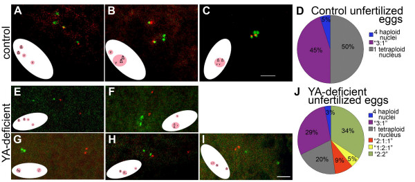
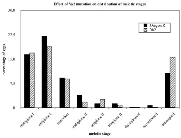
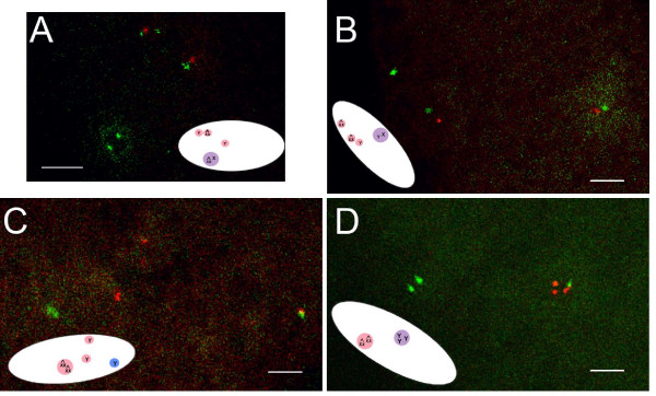
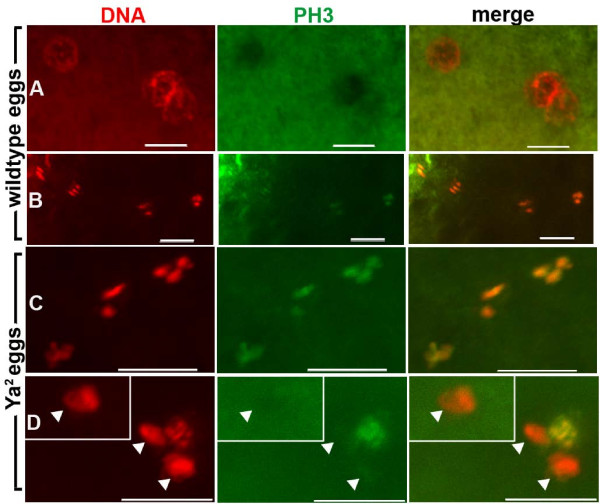
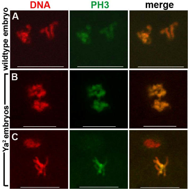
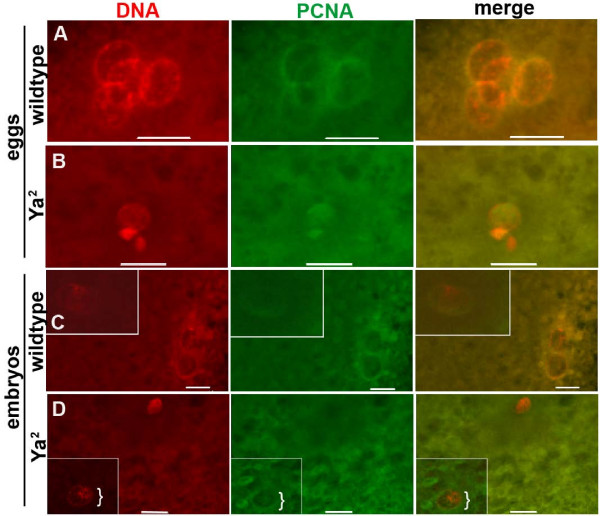
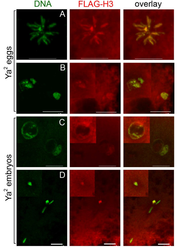
Results: We find that meiosis is completed normally in the absence of YA function. The first defects in embryos and eggs from mutant mothers first appear just after the completion of meiosis, and are seen as abnormal associations among the resultant haploid nuclei. These defects are associated with asynchronies in the cell cycle-dependent chromatin condensation state of the haploid nuclei. However, we find evidence of DNA replication in the absence of YA function.

Conclusion: Our data suggest YA function is needed at a control point, following meiosis II and the initiation of the first postmeiotic S phase, which is sensitive to the chromatin condensation state of the haploid meiotic products.


Figures

Figure 1
Figure 1
Meiotic product behavior in control and YA-deficient unfertilized eggs. In situ hybridization to the X and Y chromosomes in 0–15 minute old unfertilized eggs laid by X^X/Y, Ya+ (control) females (A-C) and X^X Ya2/Y (Ya2 mutant) females (E-I). The X chromosome probe's signal is green, and the Y chromosome's is red. The green hybridization signal to the X chromosome also recognizes the maternal Y, Ya+ in control eggs (thus the Y, Ya+ is red and green in A-C). The X probe does not recognize the maternal Y chromosome in YA-deficient eggs (thus the Y is only red in E-I). (A-C) Unfertilized eggs containing (A) four separate haploid meiotic products, (B) one triploid nucleus with two X^X chromosomes and one Y chromosome, the result of association of three meiotic products, and one haploid Y-containing nucleus. (C) all four meiotic products associated. (D) Chart of percentages of nuclear distributions observed in the 22 control eggs. (E-I) YA-deficient unfertilized eggs containing (E) four haploid meiotic products, (F) one triploid nucleus with two X^X chromosomes and one Y chromosome, reflecting three associated meiotic products, and a separate haploid Y chromosome-containing nucleus, (G) two diploid nuclei, one containing two X^X chromosomes and the other containing two Y chromosomes. H and I both contain three DAPI-stained nuclei: (H) one Y-containing haploid nucleus, one diploid nucleus containing one X^X and one Y chromosome and one haploid X^X-containing nucleus, (I) two haploid X^X chromosome-containing nuclei and one diploid nucleus containing two Y chromosomes. (J) Chart of percentages of nuclear distributions observed in the 38 Ya2 eggs. DAPI staining clearly showed the number of distinct nuclei per egg (four in A, E, two in B, F, G, one in C, three in H, I). Inset illustrations in each panel show the orientation of the egg and positions of DAPI stained nuclei inside (not drawn to scale). Bar = 4 μm for all panels.
Figure 2
Figure 2
Effect of Ya2 mutation on distribution of meiotic stages. Wildtype (Oregon R P2) and X^X, Ya2/Y (Ya2 mutant) eggs were activated and staged based on DAPI staining. Eggs with apparently condensed disorganized chromatin are grouped into the "unassigned" category, although some of the eggs in this category likely had completed meiosis. There are no statistically significant differences in the distribution of stages of wildtype vs. YA-deficient eggs as assessed by Fisher's exact test; for each of the nine categories of meiotic stages, comparison of the fraction of wildtype eggs in a stage to the fraction of YA-deficient eggs in that stage gave p values ranging from p = 1 to p = 0.061. Wildtype total n = 327 eggs. Ya2 total n = 280 eggs.
Figure 3
Figure 3
Meiotic product behavior in control and YA-deficient embryos. In situ hybridization to X and Y chromosomes in 0–15 minute old embryos from X^X/Ya+Y (control) and X^X Ya2/Y (Ya2mutant) mothers mated to X/Y, Ya+males. Inset illustrations in each panel show the orientation of the embryo and positions of DAPI stained nuclei inside (not drawn to scale). Pink circles represent maternally derived nuclei, blue circles represent paternally derived nuclei. The X chromosome probe's signal is green, and the Y chromosome probe's is red. The paternal Y chromosome was Y, Ya+, and therefore is marked with both red and green signals. (A) In the control gonomeric-stage embryo shown, the polar body nuclei (one haploid maternal X^X chromosome-containing nucleus and two haploid maternal Y chromosome-containing nuclei) have not yet begun to associate. The gonomeric nucleus consists of a maternal X^X and paternal X chromosome, each seen as a single dot with the X probe. Control n = 28 embryos. (B-D) YA-deficient embryos with (B) two haploid X^X chromosome-containing nuclei and one haploid Y chromosome-containing nucleus (all of maternal origin), and one diploid nucleus which is a product of an association between a male Ya+Y chromosome-containing pronucleus and a haploid X^X chromosome-containing nucleus of maternal origin, (C) one diploid X^X-containing nucleus (of maternal origin), two haploid Y-containing nuclei (of maternal origin) and a Ya+Y containing male pronucleus, (D) one diploid X^X chromosome-containing nucleus (of maternal origin) and one triploid nucleus which is presumably a product of an association between a diploid Y chromosome-containing nucleus (or two Y-containing haploid nuclei) of maternal origin and a Ya+Y chromosome-containing male pronucleus. Ya2 n = 54 embryos. Bars = 4 μm for all panels.
Figure 4
Figure 4
Phospho-histone H3 distribution on chromatin of unfertilized wildtype and YA-deficient eggs. Immunostaining of 0–15 minute old laid unfertilized wildtype and Ya2 eggs (from Oregon R P2 and X^X Ya2/Y mothers, respectively). DNA shown in red, phospho-histone H3 (PH3) in green. A-C are projections of multiple confocal images. (A) Four decondensed wildtype nuclei in promeiotic interphase or S phase, with PH3 excluded from nuclei (any faint PH3 staining was around the nuclear periphery). (B) Four condensed wildtype meiotic products perpendicular to the egg cortex, with PH3 staining on all chromatin. (C) Condensed Ya2 meiotic products (probably 1:1:2, post meiotic but just after telophase since still arranged linearly), with PH3 staining on all chromatin. (D) Four Ya2 meiotic products in two planes; three grouped at the egg cortex and one deeper in (inset). Although all four nuclei have similar nuclear areas, and thus are likely all haploid, only one of the three haploid nuclei at the cortex forming the polar body has individualized chromosomes with PH3 staining. Arrowheads indicate decondensed nuclei without PH3 staining. Wildtype n = 64 eggs. Ya2 n = 20 eggs. Bar = 10 μm.
Figure 5
Figure 5
Phospho-histone H3 distribution on chromatin of wildtype and YA-deficient embryos. Immunostaining of 0–15 minute old wildtype and Ya2 embryos. DNA shown in red, phospho-histone H3 in green. Embryos shown had three peripheral nuclei (not shown) and two nuclei apposed in the mid-anterior region. Apposed nuclei are shown. (A) Wildtype, (B) Synchronous apposed nuclei in a Ya2 embryo (70% of Ya2 embryos with apposed nuclei showed this phenotype), (C) Asynchronous apposed nuclei in a Ya2 embryo (30% of Ya2 embryos with apposed nuclei showed this phenotype). Wildtype n = 22 embryos with apposed pronuclei. Ya2 n = 27 embryos with apposed nuclei. Bar = 10 μm.
Figure 6
Figure 6
PCNA distribution on chromatin of wildtype and YA-deficient eggs and embryos. Immunostaining of 0–15 minute old laid unfertilized wildtype and Ya2 eggs, and wildtype and Ya2 embryos. DNA shown in red, PCNA in green. A and B are projections of multiple confocal images. (A) Four decondensed wildtype egg nuclei with PCNA on DNA, likely in S phase. 55% of wildtype eggs had PCNA staining nuclei, all synchronous. (B) Variously condensed Ya2 egg meiotic products with PCNA staining on 2 out of 3 areas of chromatin. 26% of Ya2 eggs had PCNA-positive nuclei; 2% were synchronous with PCNA, 24% were asynchronous. (C) Embryo with five decondensed wildtype meiotic products with PCNA staining on all. Two apposed pronuclei (inset) and three meiotic products forming a polar body (only two are visible in this plane). (D) Ya2 embryo with two nuclei in two planes; one near the embryo cortex (inset) and one deeper in. Only one of the two has PCNA staining (bracket). Wildtype n = 42 eggs, n = 215 embryos. Ya2 n = 34 eggs, n = 168 embryos. Bar = 10 μm.
Figure 7
Figure 7
Histone H3-FLAG distribution on chromatin of YA-deficient eggs and embryos. Immunostaining of 0–15 minute old laid unfertilized H3-FLAG; Ya2 eggs, and H3-FLAG;Ya2 embryos. DNA shown in red, H3-FLAG in green. A-D are projections of multiple confocal images, with insets used to show nuclei in a greatly different focal plane. (A) YA-deficient egg with polar body rosette with H3-FLAG staining. (B) YA-deficient egg with four female meiotic products all with H3-FLAG staining. (C) YA-deficient embryo with two apposed nuclei and a third nearer the egg cortex, all with H3-FLAG staining. (D) YA-deficient embryo with five nuclei, two of which are more rounded and have H3-FLAG staining while three others do not. The H3-FLAG-staining nucleus not in the inset is closer to the three unstained nuclei along the Z axis than it is to the other H3-FLAG-staining nucleus shown in the inset. Ya2 eggs n = 24, embryos n = 58. Bar = 10 μm.

Similar articles

Cited by

References

    1. Heifetz Y, Yu J, Wolfner MF. Ovulation triggers activation of Drosophila oocytes. Dev Biol. 2001;234:416–24. - PubMed
    1. Mahowald AP, Goralski TJ, Caulton JH. In vitro activation of Drosophila eggs. Dev Biol. 1983;98:437–45. - PubMed
    1. Page AW, Orr-Weaver TL. Activation of the meiotic divisions in Drosophila oocytes. Dev Biol. 1997;183:195–207. - PubMed
    1. Horner VL, Czank A, Jang JK, Singh N, Williams BC, Puro J, Kubli E, Hanes SD, McKim KS, Wolfner MF, Goldberg ML. The Drosophila calcipressin sarah is required for several aspects of egg activation. Curr Biol. 2006;16:1441–6. - PubMed
    1. Takeo S, Tsuda M, Akahori S, Matsuo T, Aigaki T. The calcineurin regulator sra plays an essential role in female meiosis in Drosophila. Curr Biol. 2006;16:1435–40. - PubMed

Publication types

MeSH terms

Substances

LinkOut - more resources