

Gastroesophageal reflux leads to esophageal cancer in a surgical model with mice
- PMID: 19627616
- PMCID: PMC2723127
- DOI: 10.1186/1471-230X-9-59
Gastroesophageal reflux leads to esophageal cancer in a surgical model with mice
Abstract
Background: Esophago-gastroduodenal anastomosis with rats mimics the development of human Barrett's esophagus and esophageal adenocarcinoma by introducing mixed reflux of gastric and duodenal contents into the esophagus. However, use of this rat model for mechanistic and chemopreventive studies is limited due to lack of genetically modified rat strains. Therefore, a mouse model of esophageal adenocarcinoma is needed.
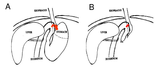
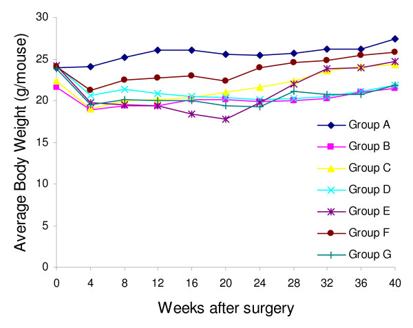
Methods: We performed reflux surgery on wild-type, p53A135V transgenic, and INK4a/Arf+/- mice of A/J strain. Some mice were also treated with omeprazole (1,400 ppm in diet), iron (50 mg/kg/m, i.p.), or gastrectomy plus iron. Mouse esophagi were harvested at 20, 40 or 80 weeks after surgery for histopathological analysis.
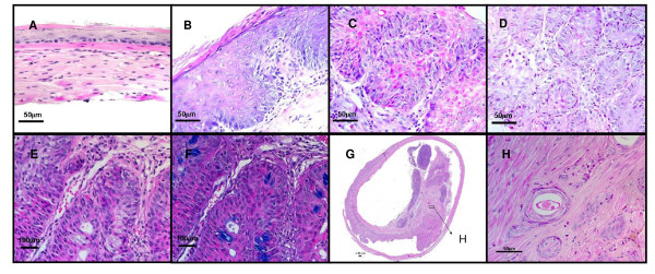
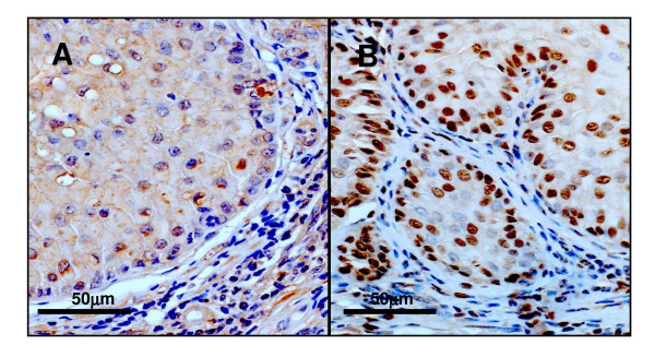
Results: At week 20, we observed metaplasia in wild-type mice (5%, 1/20) and p53A135V mice (5.3%, 1/19). At week 40, metaplasia was found in wild-type mice (16.2%, 6/37), p53A135V mice (4.8%, 2/42), and wild-type mice also receiving gastrectomy and iron (6.7%, 1/15). Esophageal squamous cell carcinoma developed in INK4a/Arf+/- mice (7.1%, 1/14), and wild-type mice receiving gastrectomy and iron (21.4%, 3/14). Among 13 wild-type mice which were given iron from week 40 to 80, twelve (92.3%) developed squamous cell carcinoma at week 80. None of these mice developed esophageal adenocarcinoma.
Conclusion: Surgically induced gastroesophageal reflux produced esophageal squamous cell carcinoma, but not esophageal adenocarcinoma, in mice. Dominant negative p53 mutation, heterozygous loss of INK4a/Arf, antacid treatment, iron supplementation, or gastrectomy failed to promote esophageal adenocarcinoma in these mice. Further studies are needed in order to develop a mouse model of esophageal adenocarcinoma.
Figures
Similar articles
-
Influence of surgically induced gastric and gastroduodenal content reflux on esophageal carcinogenesis--experimental model in Wistar female rats.Dis Esophagus. 1999;12(2):106-15. doi: 10.1046/j.1442-2050.1999.00011.x. Dis Esophagus. 1999. PMID: 10466042
-
Reflux of duodenal or gastro-duodenal contents induces esophageal carcinoma in rats.Int J Cancer. 1996 Jul 17;67(2):269-74. doi: 10.1002/(SICI)1097-0215(19960717)67:2<269::AID-IJC19>3.0.CO;2-6. Int J Cancer. 1996. PMID: 8760598
-
Animal Model: Reflux Models in Esophageal Adenocarcinoma.Methods Mol Biol. 2018;1756:143-150. doi: 10.1007/978-1-4939-7734-5_13. Methods Mol Biol. 2018. PMID: 29600367
-
[Reflux of duodenal or gastroduodenal contents induces esophageal carcinoma in rats].Nihon Rinsho. 2004 Aug;62(8):1433-8. Nihon Rinsho. 2004. PMID: 15344531 Review. Japanese.
-
Rat Reflux Model of Esophageal Cancer and Its Implication in Human Disease.Ann Surg. 2015 Dec;262(6):910-24. doi: 10.1097/SLA.0000000000001207. Ann Surg. 2015. PMID: 25822684 Review.
Cited by
-
Correlation Analysis of circRNA Circ_0071662 in Diagnosis and Prognosis of Esophageal Squamous Cell Carcinoma.Int J Gen Med. 2021 Dec 29;14:10423-10428. doi: 10.2147/IJGM.S343889. eCollection 2021. Int J Gen Med. 2021. PMID: 35002298 Free PMC article.
-
Development and characterization of a surgical mouse model of reflux esophagitis and Barrett's esophagus.J Gastrointest Surg. 2014 Feb;18(2):234-40; discussion 240-1. doi: 10.1007/s11605-013-2386-z. Epub 2013 Nov 5. J Gastrointest Surg. 2014. PMID: 24190247
-
Transitional basal cells at the squamous-columnar junction generate Barrett's oesophagus.Nature. 2017 Oct 26;550(7677):529-533. doi: 10.1038/nature24269. Epub 2017 Oct 12. Nature. 2017. PMID: 29019984 Free PMC article.
-
Review: Experimental models for Barrett's esophagus and esophageal adenocarcinoma.Am J Physiol Gastrointest Liver Physiol. 2012 Jun 1;302(11):G1231-43. doi: 10.1152/ajpgi.00509.2011. Epub 2012 Mar 15. Am J Physiol Gastrointest Liver Physiol. 2012. PMID: 22421618 Free PMC article. Review.
-
Barrett's Metaplasia Progression towards Esophageal Adenocarcinoma: An Attempt to Select a Panel of Molecular Sensors and to Reflect Clinical Alterations by Experimental Models.Int J Mol Sci. 2022 Mar 18;23(6):3312. doi: 10.3390/ijms23063312. Int J Mol Sci. 2022. PMID: 35328735 Free PMC article.
References
Publication types
MeSH terms
Substances
Grants and funding
LinkOut - more resources
Full Text Sources
Other Literature Sources
Medical
Molecular Biology Databases
Research Materials
Miscellaneous
