

Review
doi: 10.1016/j.pmrj.2009.02.013.
Mesenchymal stem cells: emerging therapy for Duchenne muscular dystrophy
Affiliations
- PMID: 19627945
- PMCID: PMC2746358
- DOI: 10.1016/j.pmrj.2009.02.013
Item in Clipboard
Review
Mesenchymal stem cells: emerging therapy for Duchenne muscular dystrophy
PM R.
2009 Jun.
Abstract
Multipotent cells that can give rise to bone, cartilage, fat, connective tissue, and skeletal and cardiac muscle are termed mesenchymal stem cells. These cells were first identified in the bone marrow, distinct from blood-forming stem cells. Based on the embryologic derivation, availability, and various pro-regenerative characteristics, research exploring their use in cell therapy shows great promise for patients with degenerative muscle diseases and a number of other conditions. In this review, the authors explore the potential for mesenchymal stem cell therapy in the emerging field of regenerative medicine with a focus on treatment for Duchenne muscular dystrophy.
Figures
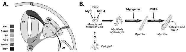
A. In the developing embryo, muscle formation is regulated by signaling pathways on either side of the notochord (NC). Within the somite (S) are the sclerotome and dermomyotome (DM). Signals from the notochord, neural tube (NT) and surface ectoderm (SE) begin events that lead to myogenic differentiation. The ventral neural tube and notochord produce Sonic hedgehog (Shh), whereas the dorsal neural tube produces Wnt-1. B. Pax-3, Myf-5 and MRF4 (Myf-6) activate MyoD in mesodermal precursor cells committing them to the myogenic lineage.
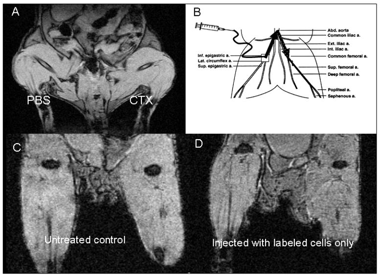
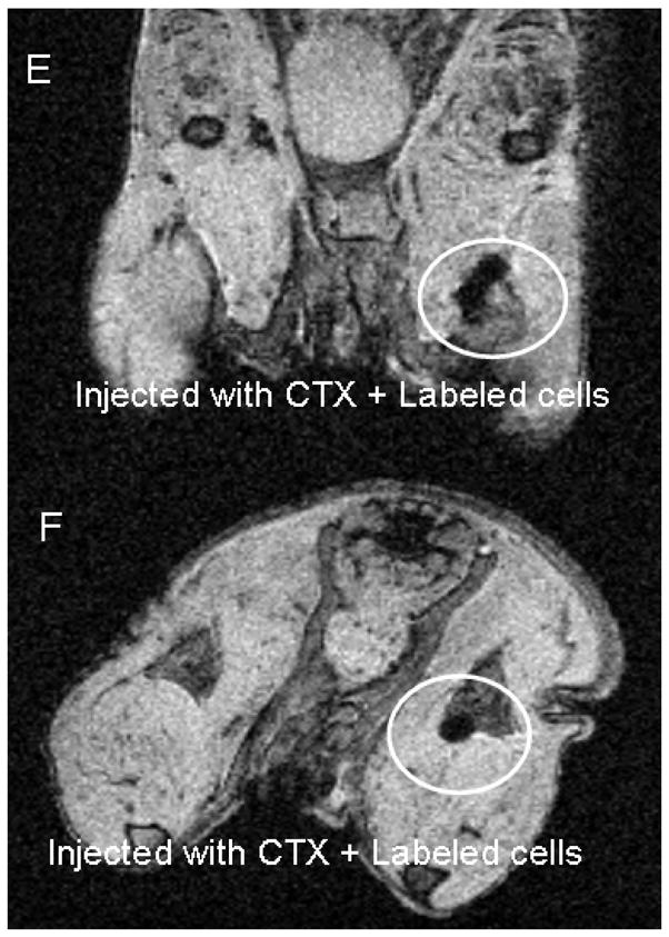
A. MRI of mouse following intramuscular injection of phosphate-buffered saline (PBS) or cadiotoxin (CTX) B. Femoral artery injection method in mice. Square indicates catheter insertion site, solid arrow indicates catheter path, dotted arrow indicates path of injected cells. C. Untreated control mouse. D. Mouse injected with iron oxide-labeled perivascular stem cells. E. and F. Mouse injected first with CTX, then with labeled cells. Circled area indicates region of labeled cells. Coronal and axial views shown in E and F, respectively.
A. MRI of mouse following intramuscular injection of phosphate-buffered saline (PBS) or cadiotoxin (CTX) B. Femoral artery injection method in mice. Square indicates catheter insertion site, solid arrow indicates catheter path, dotted arrow indicates path of injected cells. C. Untreated control mouse. D. Mouse injected with iron oxide-labeled perivascular stem cells. E. and F. Mouse injected first with CTX, then with labeled cells. Circled area indicates region of labeled cells. Coronal and axial views shown in E and F, respectively.
Similar articles
-
Co-Transplantation of Bone Marrow-MSCs and Myogenic Stem/Progenitor Cells from Adult Donors Improves Muscle Function of Patients with Duchenne Muscular Dystrophy.Cells. 2020 Apr 30;9(5):1119. doi: 10.3390/cells9051119. Cells. 2020. PMID: 32365922 Free PMC article.
-
[Treatment progress of Duchenne Muscular Dystrophy (DMD)].Med Wieku Rozwoj. 2004 Jan-Mar;8(1):25-32. Med Wieku Rozwoj. 2004. PMID: 15557694 Review. Polish.
-
Transplantation of Dystrophin Expressing Chimeric Human Cells of Myoblast/Mesenchymal Stem Cell Origin Improves Function in Duchenne Muscular Dystrophy Model.Stem Cells Dev. 2021 Feb;30(4):190-202. doi: 10.1089/scd.2020.0161. Epub 2021 Jan 22. Stem Cells Dev. 2021. PMID: 33349121
-
Perspectives of stem cell therapy in Duchenne muscular dystrophy.FEBS J. 2013 Sep;280(17):4251-62. doi: 10.1111/febs.12083. Epub 2013 Jan 7. FEBS J. 2013. PMID: 23206279 Review.
-
Immunomodulatory amnion-derived mesenchymal stromal cells preserve muscle function in a mouse model of Duchenne muscular dystrophy.Stem Cell Res Ther. 2023 Apr 27;14(1):108. doi: 10.1186/s13287-023-03337-0. Stem Cell Res Ther. 2023. PMID: 37106393 Free PMC article.
Cited by
-
Photoacoustic imaging of mesenchymal stem cells in living mice via silica-coated gold nanorods.ACS Nano. 2012 Jul 24;6(7):5920-30. doi: 10.1021/nn302042y. Epub 2012 Jun 20. ACS Nano. 2012. PMID: 22681633 Free PMC article.
-
In Vivo Photoacoustic Monitoring of Stem Cell Location and Apoptosis with Caspase-3-Responsive Nanosensors.ACS Nano. 2023 Sep 26;17(18):17931-17945. doi: 10.1021/acsnano.3c04161. Epub 2023 Sep 13. ACS Nano. 2023. PMID: 37703202 Free PMC article.
-
Myogenic-induced mesenchymal stem cells are capable of modulating the immune response by regulatory T cells.J Tissue Eng. 2014 Feb 18;5:2041731414524758. doi: 10.1177/2041731414524758. eCollection 2014. J Tissue Eng. 2014. PMID: 24555015 Free PMC article.
-
Cyclooxygenase-2 or tumor necrosis factor-α inhibitors attenuate the mechanotransductive effects of pulsed focused ultrasound to suppress mesenchymal stromal cell homing to healthy and dystrophic muscle.Stem Cells. 2015 Apr;33(4):1173-86. doi: 10.1002/stem.1927. Stem Cells. 2015. PMID: 25534849 Free PMC article.
-
Coaxing stem cells for skeletal muscle repair.Adv Drug Deliv Rev. 2015 Apr;84:198-207. doi: 10.1016/j.addr.2014.07.007. Epub 2014 Jul 15. Adv Drug Deliv Rev. 2015. PMID: 25049085 Free PMC article. Review.
References
Publication types
MeSH terms
Grants and funding
LinkOut - more resources
Full Text Sources
Other Literature Sources
