Comparative Study
. 2009 Oct 15;334(2):523-34.
doi: 10.1016/j.ydbio.2009.07.012. Epub 2009 Jul 21.

Transcription of the C. elegans let-7 microRNA is temporally regulated by one of its targets, hbl-1

Affiliations
Comparative Study

Transcription of the C. elegans let-7 microRNA is temporally regulated by one of its targets, hbl-1

Sarah F Roush et al. Dev Biol. .

Abstract

The let-7 family of microRNAs (miRNAs) are important regulators of developmental timing and cell differentiation and are often misexpressed in human cancer. In C. elegans, let-7 controls cell fate transitions from larval stage 4 (L4) to adulthood by post-transcriptionally down-regulating lineage-abnormal 41 (lin-41) and hunchback-like 1 (hbl-1). Primary let-7 (pri-let-7) transcripts are up-regulated in the L3, yet little is known about what controls this transcriptional up-regulation. We sought factors that either turn on let-7 transcription or keep it repressed until the correct time. Here we report that one of let-7's targets, the transcription factor Hunchback-like 1 (HBL-1), is responsible for inhibiting the transcription of let-7 in specific tissues until the L3. hbl-1 is a known developmental timing regulator and inhibits adult development in larval stages. Therefore, one important function of HBL-1 in maintaining larval stage fates is inhibition of let-7. Indeed, our results reveal let-7 as the first known target of the HBL-1 transcription factor in C. elegans and suggest a negative feedback loop mechanism for let-7 and HBL-1 regulation.


Figures
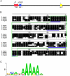
Figure 1
Figure 1
The let-7 promoter contains a 62 bp region of high conservation wherein lie three putative HBL-1 binding sites. (A) Schematic of the C. elegans let-7 promoter showing the positions of TRE and putative HBL-1 binding sites. Red box: Temporal Regulatory Region (TRE); blue box: 62 bp region containing putative HBL-1 binding sites (HBL-1); yellow box: mature let-7. (B) Sequence alignment of region containing putative HBL-1 binding sites in the let-7 promoter from 4 nematode species. Green outlines the 62 bp element. Potential HBL-1 binding sites are underlined in blue. These sites do show variation with the Drosophila melanogaster Hunchback consensus binding site. (C) Drosophila melanogaster Hunchback consensus binding site from the TESS database (Schug, 1997).
Figure 2
Figure 2
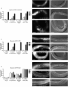
Deletion of the 62 bp region from the let-7::gfp transcriptional reporter causes precocious and sustained GFP expression in transgenic lines. (A,F,K) N ≥ 30 animals for each stage. (A) GFP is expressed precociously in the seam cells of animals where the 62 bp element has been deleted from the reporter construct (Δhbl-1::gfp). (B) GFP image of an L1 let-7::gfp animal showing that there is no GFP expression in the seam cells. (C) Nomarski DIC image showing the stage of the animal in (B). (D) GFP image of an L1 Δhbl-1::gfp animal showing expression in the seam cells, as indicated by arrows. (E) Nomarski DIC image showing the stage of the animal in (D). (F) GFP is expressed precociously in the VPCs of Δhbl-1::gfp animals. (G) GFP image of an L2 let-7::gfp animal showing that there is no GFP expression in the VPCs. (H) Nomarski DIC image showing the stage of the animal in (G). (I) GFP image of an L2 Δhbl-1::gfp animal showing expression in the VPCs, as indicated by arrows. (J) Nomarski DIC image showing the stage of the animal in (I). (K) GFP expression persists in later larval stages in Δhbl-1::gfp animals. (L) GFP image of an mL3 let-7::gfp animal showing no GFP expression in the hyp7, indicated by brackets, while there is expression in the seam cells, indicated by asterisks. (M) Nomarski DIC image showing the stage of the animal in (L). (N) GFP and Nomarski DIC images of an mL3 Δhbl-1::gfp animal showing expression in the hyp7, as indicated by brackets. Asterisks indicate seam cells. Lack of GFP expression in all of the seam cells may be due to mosaicism in the transgenic line. (O) Nomarski DIC image showing the stage of the animal in (N). Scale bars all represent 20 microns. Anterior is left and dorsal is top on all images.
Figure 3
Figure 3
Deletion of the 62 bp region from the let-7 rescue fragment causes an increase in precocious alae expression. (A) At the eL4 stage, wild-type (N2) animals do not express alae, while wild-type animals injected with the wild-type rescue fragment express precocious alae at a low level. Significantly more wild-type animals injected with a rescue fragment that has the 62 bp region deleted show precocious alae. n ≥ 30 for each strain. Three independent lines were analyzed for each construct. (B) Nomarski DIC images of wild-type eL4 C. elegans showing that no alae are expressed. (C) Nomarski DIC image showing the stage of the animal in (B). (D) Nomarski DIC images of wild-type eL4 C. elegans injected with the let-7 rescue fragment which has the 62 bp region deleted showing precociously expressed alae. Arrows indicate alae. (E) Nomarski DIC image showing the stage of the animal in (D). (F) Wild-type eL4 animal fed 50% hbl-1(RNAi) which shows precocious alae formation similar to precocious alae seen in C. elegans injected with the 62 bp deleted rescue fragment. Arrows indicate alae. Precocious alae in hbl-1(lf) has previously been identified in other studies (Abrahante et al., 2003; Lin et al., 2003). (G) Nomarski DIC image showing the stage of the animal in (F). Scale bars all represent 20 microns. Anterior is left and dorsal is top on all images.
Figure 4
Figure 4
Reduced HBL-1 activity causes precocious and sustained let-7::gfp expression similar to Δhbl1::gfp. (A,H,O) N ≥ 30 animals for each stage. (A) When let-7::gfp is crossed with the hbl-1(ve18) loss-of-function mutant or grown on 50% hbl-1(RNAi), there is precocious Δhbl1::gfp expression in the seam cells beginning in L1. (B) GFP image of an L2 let-7::gfp animal showing that there is no GFP expression in the seam cells. (C) Nomarski DIC image showing the stage of the animal in (B). (D) GFP image of an L2 let-7::gfp;hbl-1(ve18) animal showing expression in the seam cells, as indicated by arrows. (E) Nomarski DIC image showing the stage of the animal in (D). (F) GFP image of an L2 let-7::gfp;50% hbl-1(RNAi) animal showing expression in the seam cells, as indicated by arrows. (G) Nomarski DIC image showing the stage of the animal in (F). (H) When let-7::gfp is crossed with the hbl-1(ve18) loss-of-function mutant or grown on 50% hbl-1(RNAi), there is precocious let-7::gfp expression in the VPCs beginning in L2 or eL3, respectively. (I) GFP image of an eL3 let-7::gfp animal showing that there is no GFP expression in the VPCs. (I) Nomarski DIC image showing the stage of the animal in (J). (K) GFP image of an eL3 let-7::gfp;hbl-1(ve18) animal showing expression in the VPCs, as indicated by arrows. (L) Nomarski DIC image showing the stage of the animal in (K). (M) GFP image of an eL3 let-7::gfp;50% hbl-1(RNAi) animal showing expression in the VPCs, as indicated by arrows. (N) Nomarski DIC image showing the stage of the animal in (M). (O) let-7::gfp expression is sustained in the hyp7 in a higher proportion of animals through later larval stages in hbl-1(ve18) loss-of-function mutant or grown on 50% hbl-1(RNAi). It is unknown why there is an increase in GFP detected between let-7::gfp C. elegans fed wild-type bacteria versus bacteria expressing the RNAi empty vector, L4440. Importantly, the same pattern of increased GFP detection was seen when hbl-1 levels were reduced by mutation or RNAi. (P) GFP image of an mL3 let-7::gfp animal showing no GFP expression in the hyp7, indicated by brackets, while there is expression in the seam cells, indicated by asterisks. (Q) Nomarski DIC image showing the stage of the animal in (P). (R,S) GFP image of an mL3 let-7::gfp;hbl-1(ve18) animal showing expression in the hyp7, as indicated by brackets. Seam cells are indicated by asterisks. (S) Nomarski DIC image showing the stage of the animal in (R). (T) GFP image of an mL3 let-7::gfp;50% hbl-1(RNAi) animal showing expression in the hyp7, as indicated by brackets. Seam cells are indicated by asterisks. (U) Nomarski DIC image showing the stage of the animal in (T). Scale bars all represent 20 microns. Anterior is left and dorsal is top on all images.
Figure 5
Figure 5
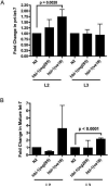
Levels of SL1-spliced pri-let-7 and mature let-7 are increased in hbl-1 loss-of-function mutants when measured by qRT-PCR. (A) The amount of SL1-spliced pri-let-7 during the L2 stage was significantly increased by approximately 75% in the hbl-1(ve18) mutant. The hbl-1(mg285) mutant also showed an increase in the amount of SL1-spliced pri-let-7 present in the L2 stage, but this value was not significant. The SL1-spliced pri-let-7 levels in the hbl-1 mutants return to levels similar to those detected in wild-type C. elegans during the L3 stage. The standard deviations are shown. (B) Levels of mature let-7 are significantly increased by approximately 2-fold during the L3 stage in the hbl-1(ve18) mutant. The increase in let-7 in hbl-1(ve18) in the L2 stage is not statistically significant. The standard deviations are shown. Paired t-tests were used to measure statistical significance. Three technical replicates from two independent biological replicates were assayed for each column in Fig. 5.
Figure 6
Figure 6
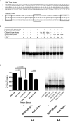
A probe using the 62 bp region containing putative HBL-1 binding sites is specifically shifted in EMSAs by wild-type C. elegans protein extracts, and this shift is significantly decreased when extracts from hbl-1 loss-of-function mutants and 50% hbl-1(RNAi) are used. (A) Wild-type and mutant probe sequences used in EMSAs. Boxed sequences in the mutant probe indicate the mutations and location of potential HBL-1 sites. (B) Lanes 1−4, the wild-type probe, but not a probe with mutated HBL-1 binding sites, is shifted by C. elegans protein extract. Lanes 5−8, increasing amount of non-radiolabeled, wild-type probe (10, 50 and 100× greater than labeled probe) can compete away the labeled probe's shift. Lanes 9−12, increasing amounts of non-radiolabeled mutant probe cannot compete away wild-type probe's shift. (C) EMSAs using protein extracts from hbl-1(ve18), hbl-1(mg285) and 50% hbl-1(RNAi), have a significant decrease in the amount of probe shifted. n = 5, a representative gel is shown. Paired t-tests were used to measure statistical significance.

Similar articles

Cited by

References

    1. Abbott AL, Alvarez-Saavedra E, Miska EA, Lau NC, Bartel DP, Horvitz HR, Ambros V. The let-7 MicroRNA family members mir-48, mir-84, and mir-241 function together to regulate developmental timing in Caenorhabditis elegans. Dev Cell. 2005;9:403–14. - PMC - PubMed
    1. Abrahante JE, Daul AL, Li M, Volk ML, Tennessen JM, Miller EA, Rougvie AE. The Caenorhabditis elegans hunchback-like gene lin-57/hbl-1 controls developmental time and is regulated by microRNAs. Dev Cell. 2003;4:625–37. - PubMed
    1. Ambros V. A hierarchy of regulatory genes controls a larva-to-adult developmental switch in C. elegans. Cell. 1989;57:49–57. - PubMed
    1. Baskerville S, Bartel DP. Microarray profiling of microRNAs reveals frequent coexpression with neighboring miRNAs and host genes. Rna. 2005;11:241–7. - PMC - PubMed
    1. Bohnsack MT, Czaplinski K, Gorlich D. Exportin 5 is a RanGTP-dependent dsRNA-binding protein that mediates nuclear export of pre-miRNAs. Rna. 2004;10:185–91. - PMC - PubMed

Publication types

MeSH terms

LinkOut - more resources