. 2009 Sep 1;391(2):171-6.
doi: 10.1016/j.virol.2009.06.045. Epub 2009 Jul 22.

RIG-I is cleaved during picornavirus infection

Affiliations

RIG-I is cleaved during picornavirus infection

Paola M Barral et al. Virology. .

Abstract

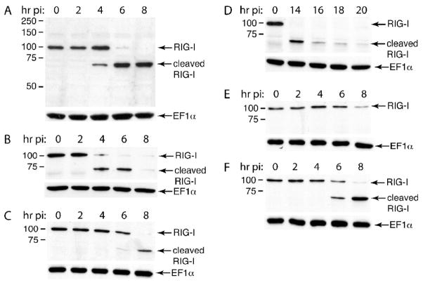
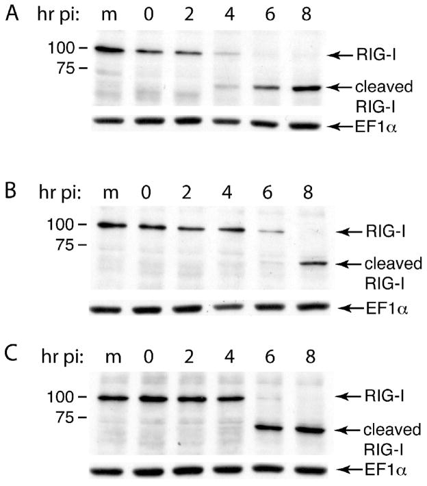
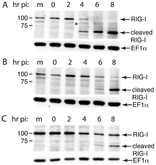
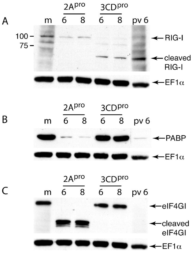
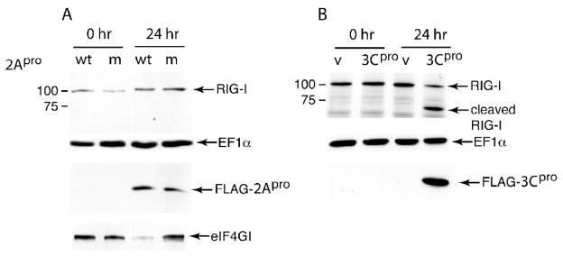
The innate immune system senses RNA virus infections through membrane-bound Toll-like receptors or the cytoplasmic proteins RIG-I and MDA-5. RIG-I is believed to recognize the 5'-triphosphate present on many viral RNAs, and hence is important for sensing infections by paramyxoviruses, influenza viruses, rhabdoviruses, and flaviviruses. MDA-5 recognizes dsRNA, and senses infection with picornaviruses, whose RNA 5'-ends are linked to a viral protein, VPg, not a 5'-triphosphate. We previously showed that MDA-5 is degraded in cells infected with different picornaviruses, and suggested that such cleavage might be a mechanism to antagonize production of type I IFN in response to viral infection. Here we examined the state of RIG-I during picornavirus infection. RIG-I is degraded in cells infected with poliovirus, rhinoviruses, echovirus, and encephalomyocarditis virus. In contrast to MDA-5, cleavage of RIG-I is not accomplished by cellular caspases or the proteasome. Rather, the viral proteinase 3C(pro) cleaves RIG-I, both in vitro and in cells. Cleavage of RIG-I during picornavirus infection may constitute another mechanism for attenuating the innate response to viral infection.


Figures

Figure 1
Figure 1. Degradation of RIG-I during picornavirus infection
Monolayers of HeLa cells (A, C–F) or SH-SY-5Y cells (B) were infected (MOI 10) with poliovirus (A, B), echovirus (C), rhinovirus type 16 (D), rhinovirus type 1A (E) or EMCV (F). At the indicated times after infection, cell extracts were prepared, fractionated by SDS-PAGE, and RIG-I was detected by western blot analysis. Positions of RIG-I and a putative cleavage product are indicated. Separate bottom panels show detection of EF1α to ensure that equal amounts of protein were applied to each lane.
Figure 2
Figure 2. Effect of protease inhibitors on poliovirus-induced cleavage of RIG-I
Monolayers of HeLa cells were infected with poliovirus (MOI 10) in the absence (A) or presence of Z-VAD-FMK (100 μM) (B) or MG132 (20 μM) (C). At the indicated times after infection, cell extracts were prepared, fractionated by SDS-PAGE, and RIG-I was detected by western blot analysis. Positions of RIG-I and a putative cleavage product are indicated. Separate bottom panels show detection of EF1α to ensure that equal amounts of protein were applied to each lane.
Figure 3
Figure 3. Effect of amino acid changes in polioviral proteinases 2Apro and 3Cpro on cleavage of RIG-I
Monolayers of HeLa cells were infected with wild-type poliovirus (A), mutant 2AproY88L (B) or mutant Se1–3C-02 (C) (MOI 10). At the indicated times after infection, cell extracts were prepared, fractionated by SDS-PAGE, and RIG-I was detected by western blot analysis. Positions of RIG-I and a putative cleavage product are indicated. Separate bottom panels show detection of EF1α to ensure that equal amounts of protein were applied to each lane.
Figure 4
Figure 4. Effect of 2Apro and 3CDpro on RIG-I in vitro
A cytoplasmic extract from HeLa cells was incubated with purified 2Apro or 3CDpro for 6 or 8 hr, fractionated by SDS-PAGE, and RIG-I was detected by western blot analysis. Cytoplasmic extracts from mock-infected HeLa cells (m) and from cells 6 hr after poliovirus infection (pv 6) were included to show the location of uncleaved and cleaved RIG-I, respectively. eIF4GI and PABP (to confirm enzyme activity of 2Apro and 3CDpro) and EF1α (loading control) were detected by western blot analysis.
Figure 5
Figure 5. Effect of 2Apro and 3Cpro on RIG-I in vivo
Plasmids encoding poliovirus 2Apro or 3Cpro (each with an N-terminal FLAG epitope) or vector alone (v) were introduced into HeLa cells by transformation. At 0 and 24 after DNA-mediated transformation, cell extracts were prepared, fractionated by SDS-PAGE, and RIG-I was detected by western blot analysis. Production of each viral proteinase was confirmed by western analysis using anti-FLAG antibody. eIF4GI (to confirm enzyme activity of 2Apro) and EF1α (loading control) were detected by western blot analysis.

Similar articles

Cited by

References

    1. Barral PM, Morrison JM, Drahos J, Gupta P, Sarkar D, Fisher PB, Racaniello VR. MDA-5 is cleaved in poliovirus-infected cells. J Virol. 2007;81(8):3677–84. - PMC - PubMed
    1. Clark ME, Hämmerle T, Wimmer E, Dasgupta A. Poliovirus proteinase 3C converts an active form of transcription factor IIIC to an inactive form: a mechanism for inhibition of host cell polymerase III transcription by poliovirus. EMBO J. 1991;10:2941–2947. - PMC - PubMed
    1. Dewalt PG, Semler BL. Site-directed mutagenesis of proteinase 3C results in a poliovirus deficient in synthesis of viral RNA polymerase. J Virol. 1987;61:2162–2170. - PMC - PubMed
    1. Duke GM, Palmenberg AC. Cloning and synthesis of infectious cardiovirus RNAs containing short, discrete poly(C) tracts. J Virol. 1989;63:1822–1826. - PMC - PubMed
    1. Gitlin L, Barchet W, Gilfillan S, Cella M, Beutler B, Flavell RA, Diamond MS, Colonna M. Essential role of mda-5 in type I IFN responses to polyriboinosinic:polyribocytidylic acid and encephalomyocarditis picornavirus. Proc Natl Acad Sci U S A. 2006;103 (22):8459–64. - PMC - PubMed

Publication types

LinkOut - more resources