

Structural basis for alpha-conotoxin potency and selectivity
- PMID: 19628399
- PMCID: PMC2754775
- DOI: 10.1016/j.bmc.2009.07.005
Structural basis for alpha-conotoxin potency and selectivity
Abstract
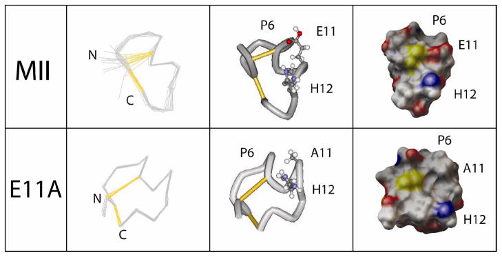
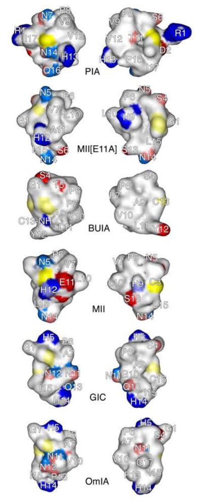
Parkinson's disease is a debilitating movement disorder characterized by altered levels of alpha(6)beta(2) * ( * indicates the possible presence of additional subunits) nicotinic acetylcholine receptors (nAChRs) localized on presynaptic striatal catecholaminergic neurons. alpha-Conotoxin MII (alpha-CTx MII) is a highly useful ligand to probe alpha(6)beta(2) nAChRs structure and function, but it does not discriminate among closely related alpha(6) * nAChR subtypes. Modification of the alpha-CTx MII primary sequence led to the identification of alpha-CTx MII[E11A], an analog with 500-5300-fold discrimination between alpha(6) * subtypes found in both human and non-human primates. alpha-CTx MII[E11A] binds most strongly (femtomolar dissociation constant) to the high affinity alpha(6) nAChR, a subtype that is selectively lost in Parkinson's disease. Here, we present the three-dimensional solution structure for alpha-CTx MII[E11A] as determined by two-dimensional (1)H NMR spectroscopy to 0.13+/-0.09A backbone and 0.45+/-0.08A heavy atom root-mean-square deviation from mean structure. Structural comparisons suggest that the increased hydrophobic area of alpha-CTx MII[E11A] relative to other members of the alpha-CTx family may be responsible for its exceptionally high affinity for alpha6alpha4beta2 * nAChR as well as discrimination between alpha(6)beta(2) and alpha(3)beta(2) containing nAChRs. This finding may enable the rational design of novel peptide analogs that demonstrate enhanced specificity for alpha(6) * nAChR subunit interfaces and provide a means to better understand nAChR structural determinants that modulate brain dopamine levels and the pathophysiology of Parkinson's disease.
Figures
References
-
- Lester HA, Dibas MI, Dahan DS, Leite JF, Dougherty DA. Trends Neurosci. 2004;27:329. - PubMed
-
- Lindstrom J. Ion Channels. 1996;4:377. - PubMed
-
- Dani JA, Bertrand D. Annu Rev Pharmacol Toxicol. 2007;47:699. - PubMed
-
- Le Novere N, Zoli M, Changeux JP. Eur J Neurosci. 1996;8:2428. - PubMed
-
- Azam L, Winzer-Serhan UH, Chen Y, Leslie FM. J Comp Neurol. 2002;444:260. - PubMed
Publication types
MeSH terms
Substances
Grants and funding
LinkOut - more resources
Full Text Sources
Chemical Information
