

Tumour-associated lymphangiogenesis in conjunctival malignant melanoma
- PMID: 19628489
- PMCID: PMC2760727
- DOI: 10.1136/bjo.2008.147355
Tumour-associated lymphangiogenesis in conjunctival malignant melanoma
Abstract
Background: To evaluate whether tumour-associated lymphangiogenesis, that is the formation of new lymphatic vessels (LVs) induced by a tumour, occurs in and around conjunctival malignant melanoma (MM).
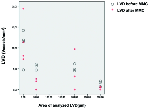
Methods: Clinical files and conjunctival specimens of 20 patients with histologically diagnosed conjunctival MM were analysed. Sections were stained with LYVE-1 and podoplanin antibodies as specific lymphatic endothelial markers and Ki67 as proliferation marker. The tumour area and the area covered by LV (LVA), LV number (LVN) and LV density (LVD) were measured within the tumour and in the peritumoural area in digital images of the specimen. The LV results were correlated with the histopathological characteristics, tumour location, recurrence rate, mitomycin C therapy and presence of metastases.
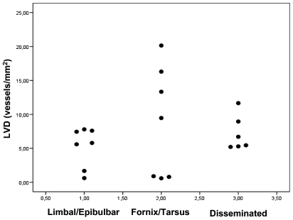
Results: LVs were detected in all specimens within the tumour and peritumourally. Significantly more Ki67(+) proliferating lymphatic endothelial cells were detected in the tumour and in the peritumoural tissue up to 300 microm compared with the surrounding normal conjunctiva (>300 microm distance). There was a slightly positive correlation between the tumour size and the LVN and LVA in the 50 microm zone adjacent to the tumour. We did not find any significant correlations between LVs and histopathological and clinical characteristics (location, shape, relapses, metastases), possibly due to the small sample sizes. Non-limbal tumours with involvement of tarsus or fornix showed a tendency towards a higher LVD compared with limbal tumours.
Conclusion: Conjunctival MMs display tumour-associated LV within and around the tumour. The MM seems to induce lymphangiogenesis not only in the tumour, but also in its proximity.
Conflict of interest statement
Figures


Similar articles
-
Prognostic significance of tumor-associated lymphangiogenesis in malignant melanomas of the conjunctiva.Ophthalmology. 2011 Dec;118(12):2351-60. doi: 10.1016/j.ophtha.2011.05.025. Epub 2011 Aug 11. Ophthalmology. 2011. PMID: 21835473
-
Tumor-associated lymphangiogenesis in the development of conjunctival squamous cell carcinoma.Ophthalmology. 2010 Apr;117(4):649-58. doi: 10.1016/j.ophtha.2010.01.032. Ophthalmology. 2010. PMID: 20346821
-
Tumor-associated lymphangiogenesis in the development of conjunctival melanoma.Invest Ophthalmol Vis Sci. 2011 Sep 9;52(10):7074-83. doi: 10.1167/iovs.11-7902. Invest Ophthalmol Vis Sci. 2011. PMID: 21849428
-
Development of lymphatic vessels: tumour lymphangiogenesis and lymphatic invasion.Curr Med Chem. 2005;12(26):3043-53. doi: 10.2174/092986705774933407. Curr Med Chem. 2005. PMID: 16375699 Review.
-
[Malignant conjunctival melanoma. Clinical review with recommendations for diagnosis, therapy and follow-up].Klin Monbl Augenheilkd. 2002 Oct;219(10):710-21. doi: 10.1055/s-2002-35693. Klin Monbl Augenheilkd. 2002. PMID: 12447715 Review. German.
Cited by
-
New Therapeutic Approaches for Conjunctival Melanoma-What We Know So Far and Where Therapy Is Potentially Heading: Focus on Lymphatic Vessels and Dendritic Cells.Int J Mol Sci. 2022 Jan 27;23(3):1478. doi: 10.3390/ijms23031478. Int J Mol Sci. 2022. PMID: 35163401 Free PMC article. Review.
-
[Adjuvant therapy and interdisciplinary follow-up care of conjunctival melanoma].Ophthalmologe. 2015 Nov;112(11):907-11. doi: 10.1007/s00347-015-0141-4. Ophthalmologe. 2015. PMID: 26502167 Review. German.
-
Conjunctival Melanoma Angiotropic Microsatellitosis: A Mechanism of Local Extravascular Migratory Metastasis.Ocul Oncol Pathol. 2020 Aug;6(4):287-292. doi: 10.1159/000505270. Epub 2020 Jan 21. Ocul Oncol Pathol. 2020. PMID: 33005619 Free PMC article.
-
[Antiangiogenic therapy at the ocular surface: when, what and why?].Ophthalmologe. 2011 Mar;108(3):230-6. doi: 10.1007/s00347-010-2262-0. Ophthalmologe. 2011. PMID: 21271256 Review. German.
-
Identification of microRNAs associated with lymphangiogenesis in human gastric cancer.Clin Transl Oncol. 2014 Apr;16(4):374-9. doi: 10.1007/s12094-013-1081-6. Epub 2013 Jul 24. Clin Transl Oncol. 2014. PMID: 23881463
References
-
- Kurli M, Finger PT. Melanocytic conjunctival tumors. Ophthalmol Clin North Am 2005;18:15–24 - PubMed
-
- Brownstein S. Malignant melanoma of the conjunctiva. Cancer Control 2004;11:310–16 - PubMed
-
- Hu DN, Yu G, McCormick SA, et al. Population-based incidence of conjunctival melanoma in various races and ethnic groups and comparison with other melanomas. Am J Ophthalmol 2008;145:418–23 - PubMed
-
- Tuomaala S, Eskelin S, Tarkkanen A, et al. Population-based assessment of clinical characteristics predicting outcome of conjunctival melanoma in whites. Invest Ophthalmol Vis Sci 2002;43:3399–408 - PubMed
Publication types
MeSH terms
Substances
LinkOut - more resources
Full Text Sources
Medical
Miscellaneous