. 2009 Nov;23(11):1850-64.
doi: 10.1210/me.2008-0260. Epub 2009 Jul 23.

A preformed signaling complex mediates GnRH-activated ERK phosphorylation of paxillin and FAK at focal adhesions in L beta T2 gonadotrope cells

Affiliations

A preformed signaling complex mediates GnRH-activated ERK phosphorylation of paxillin and FAK at focal adhesions in L beta T2 gonadotrope cells

Masha Dobkin-Bekman et al. Mol Endocrinol. 2009 Nov.

Abstract

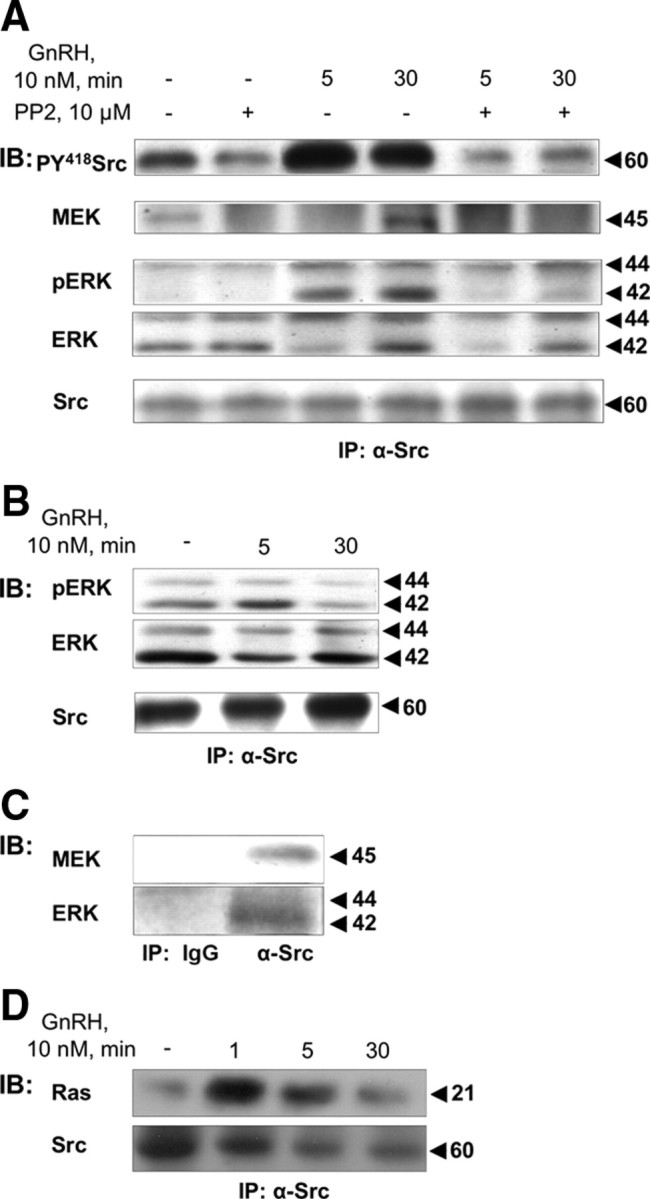
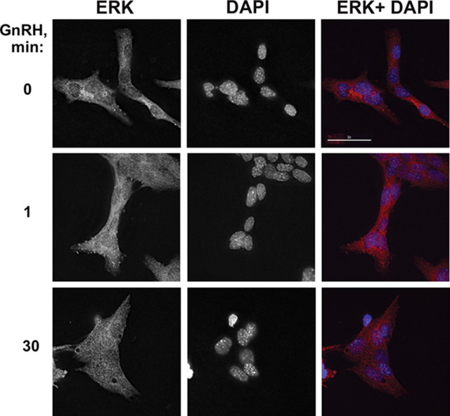
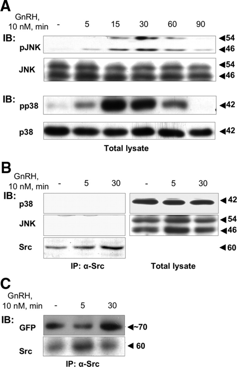
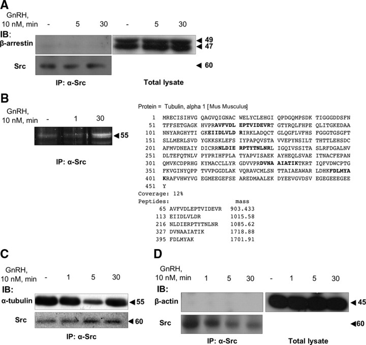
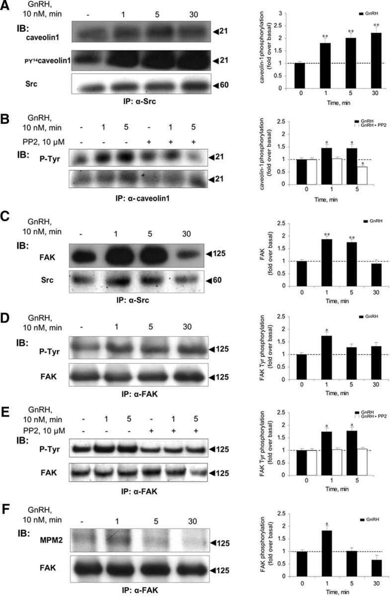
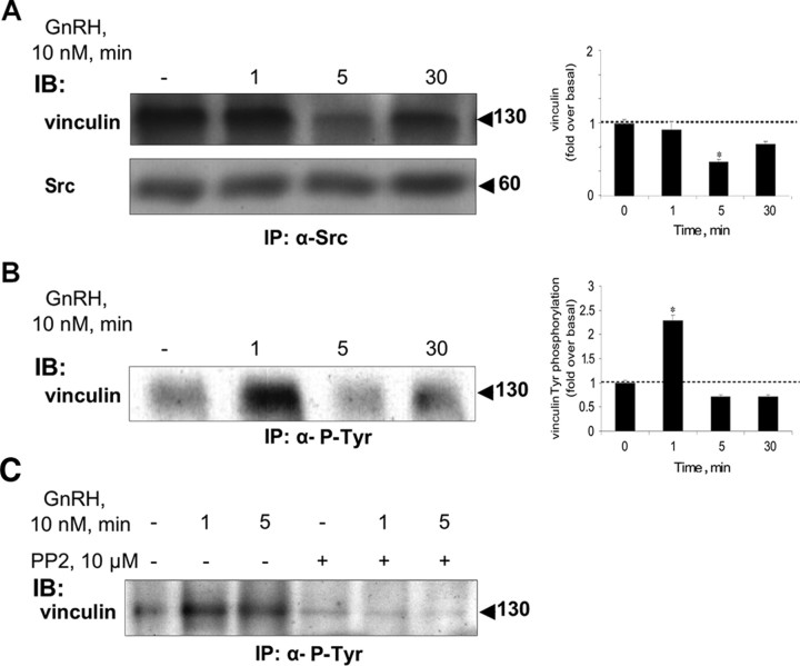
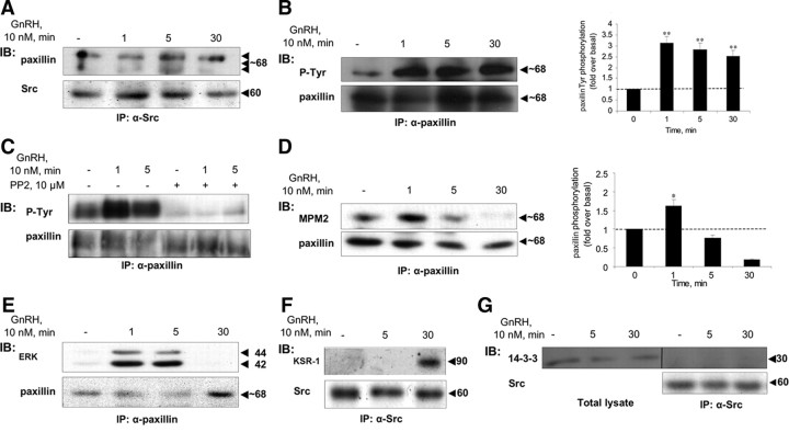
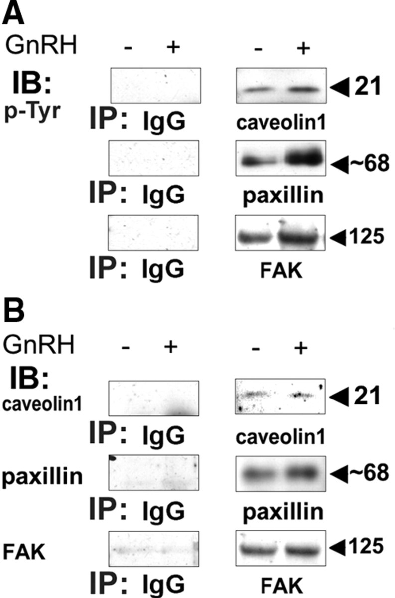
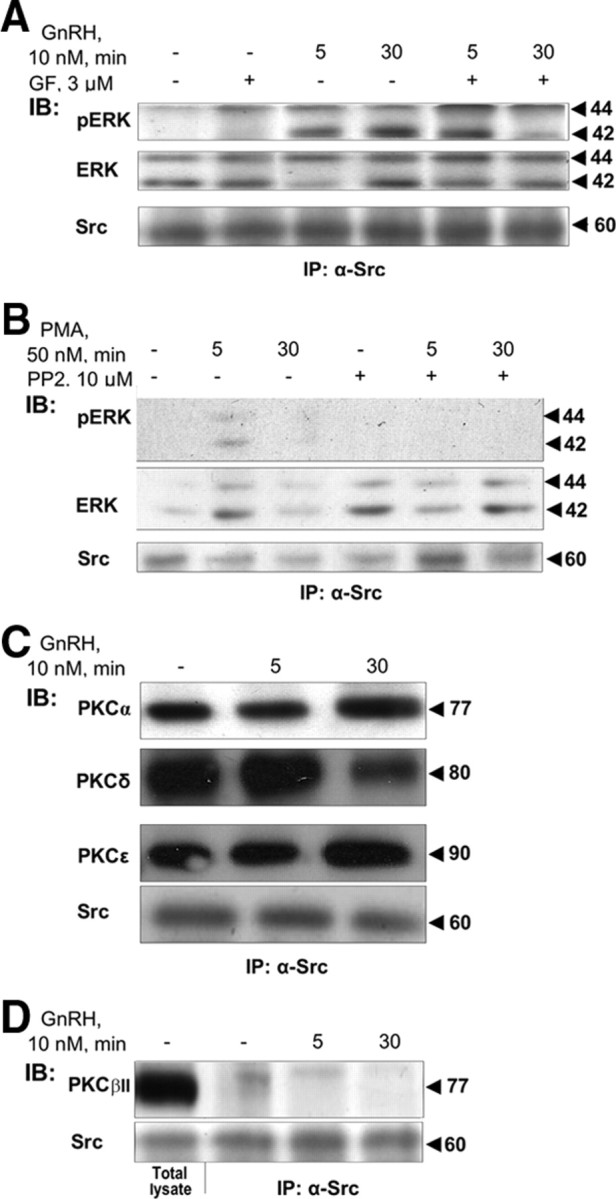
Most receptor tyrosine kinases and G protein-coupled receptors (GPCRs) operate via a limited number of MAPK cascades but still exert diverse functions, and therefore signal specificity remains an enigma. Also, most GPCR ligands utilize families of receptors for mediation of diverse biological actions; however, the mammalian type I GnRH receptor (GnRHR) seems to be the sole receptor mediating GnRH-induced gonadotropin synthesis and release. Signaling complexes associated with GPCRs may thus provide the means for signal specificity. Here we describe a signaling complex associated with the GnRHR, which is a unique GPCR lacking a C-terminal tail. Unlike other GPCRs, this signaling complex is preformed, and exposure of L beta T2 gonadotropes to GnRH induces its dynamic rearrangement. The signaling complex includes c-Src, protein kinase C delta, -epsilon, and -alpha, Ras, MAPK kinase 1/2, ERK1/2, tubulin, focal adhesion kinase (FAK), paxillin, vinculin, caveolin-1, kinase suppressor of Ras-1, and the GnRHR. Exposure to GnRH (5 min) causes MAPK kinase 1/2, ERK1/2, tubulin, vinculin, and the GnRHR to detach from c-Src, but they reassociate within 30 min. On the other hand, FAK, paxillin, the protein kinase Cs, and caveolin-1 stay bound to c-Src, whereas kinase suppressor of Ras-1 appears in the complex only 30 min after GnRH stimulation. GnRH was found to activate ERK1/2 in the complex in a c-Src-dependent manner, and the activated ERK1/2 subsequently phosphorylates FAK and paxillin. In parallel, caveolin-1, FAK, vinculin, and paxillin are phosphorylated on Tyr residues apparently by GnRH-activated c-Src. Receptor tyrosine kinases and GPCRs translocate ERK1/2 to the nucleus to phosphorylate and activate transcription factors. We therefore propose that the role of the multiprotein signaling complex is to sequester a cytosolic pool of activated ERK1/2 to phosphorylate FAK and paxillin at focal adhesions.


Figures

Fig. 1.
Fig. 1.
Activation of c-Src by GnRH and association of c-Src with Ras, MEK1/2, and ERK1/2. LβT2 cells were serum starved for 16 h before pretreatment with the c-Src inhibitor, PP2 (10 μm), for 30 min. Thereafter, GnRH (10 nm) was added for the indicated time. Cells were lysed, and proteins were subjected to SDS-PAGE after IP with α-Src antibodies. Samples were then immunoblotted (IB) with: A, antiphosphorylated Y418-Src antibodies (PY418 Src), anti-MEK antibodies, antiphospho-ERK (pERK), antitotal ERK (ERK), and anti-Src antibodies. B, Details as in panel A. Samples were IB with anti-phospho ERK (pERK), anti-total ERK (ERK), and anti-Src antibodies. C, Cells were lysed, and proteins were subjected to SDS-PAGE after IP with serum IgG, or α-Src antibodies. Samples were then IB with anti-MEK antibodies, or antitotal ERK (ERK). A representative blot is shown, and similar results were observed in two other experiments. D, LβT2 cells were serum starved for 16 h before treatment with GnRH (10 nm) for the indicated time. Cells were then lysed and subjected to SDS-PAGE after IP with α-Src antibodies and immunoblotting with specific anti-pan-Ras and anti-Src antibodies. Molecular mass (kDa) of the proteins are indicated on the right. A representative blot is shown, and similar results were observed in two other experiments.
Fig. 2.
Fig. 2.
Localization of ERK upon GnRH stimulation. LβT2 cells were serum starved overnight and were stimulated with 10 nm GnRH for the indicated periods of time. The cells were then fixed and stained with antitotal ERK antibody and DAPI as described in Materials and Methods. Removal of the secondary antibody or preabsorption of the antibodies with the relevant peptide antigen resulted in disappearance or a marked reduction of the staining, respectively (data not shown). A representative image is shown, and similar results were observed in two other experiments. Bar, 30 μm.
Fig. 3.
Fig. 3.
The c-Src immune complex is selective for ERK1/2. LβT2 cells were serum starved for 16 h before treatment with GnRH (10 nm) for the indicated time. Cells were lysed and subjected to SDS-PAGE directly (Total lysate) or after IP with α-Src antibodies (B and C). A, Activation pattern of JNK and p38MAPK as seen by Western blotting with antiphospho JNK (pJNK), or antiphospho p38 (pp38) antibodies. Total JNK and p38 were detected with polyclonal antibodies as a control for sample loading. B, p38 and JNK are in the total lystae but not in complex with Src. Total lysate or immunoprecipitated samples as above were probed with anti-JNK or anti-p38 antibodies. C, GnRHR associates with the complex. LβT2 cells were transfected with GnRHR-GFP as described in Materials and Methods, serum starved for 16 h before treatment with GnRH (10 nm) for the indicated time. Cells were lysed and subjected to SDS-PAGE after IP with α-Src antibodies. Immunoblotting (IB) was performed with anti-GFP antibodies. Molecular mass (kDa) of the proteins is indicated on the right. A representative blot is shown, and similar results were observed in two other experiments.
Fig. 4.
Fig. 4.
α-Tubulin is present, but neither β-actin nor β-arrestin is found in the c-Src immune complex. LβT2 cells were serum starved for 16 h before treatment with GnRH (10 nm) for the indicated time. Cells were then lysed and subjected to SDS-PAGE directly (Total lysate), or after IP with α-Src antibodies. A, β-Arrestin is in the total lystae but not in the c-Src immune complex, as revealed by immunoblots (IB) of the c-Src immune complex with specific anti-β-arrestin antibodies. B, α-Tubulin is a binding partner in the GnRH-induced c-Src immune complex, as revealed by a proteomic analysis. SDS-PAGE of the c-Src immune complex was stained with Sypro Ruby, and a major GnRH-stimulated band was subjected to MALDI-TOF mass spectrometry. Peptide coverage map of the specific identified protein, α-tubulin, is shown. C, α-Tubulin is present in the c-Src immune complex, as revealed by IB of the c-Src immune complex with specific anti-α-tubulin antibodies. D, β-Actin is in the total lysate but not in the c-Src immune complex, as revealed by IB of the c-Src immune complex with specific anti-β-actin antibodies. Molecular mass (kDa) of the proteins is indicated on the right. A representative blot is shown, and similar results were observed in two other experiments.
Fig. 5.
Fig. 5.
Caveolin-1 and FAK are present in the c-Src immune complex. LβT2 cells were serum starved for 16 h before treatment with GnRH (10 nm) for the indicated time. Another batch of the cells was preincubated with the c-Src inhibitor, PP2 (10 μm for 30 min), followed by GnRH stimulus for the indicated time. Cells were then lysed and subjected to SDS-PAGE after IP with the appropriate antibodies. A, Caveolin-1 is present in the c-Src immune complex, as revealed by immunoblots (IB) of the c-Src immune complex with specific anticaveolin-1 antibodies (first row). GnRH induces rapid phosphorylation of Tyr14 in caveolin-1 (n = 3; P < 0.02) in the c-Src immune complex, as revealed by IB of the c-Src immune complex with anti-PY14-caveolin-1 antibodies (second row). Blotting with α-Src antibodies is shown (third row). B, The c-Src inhibitor, PP2, markedly reduced the tyrosine phosphorylation of caveolin-1. Cells were treated as above, then lysed, and subjected to SDS-PAGE after IP with α-caveolin-1 antibodies, followed by IB with anti-p-Tyr antibodies. C, FAK is present in the c-Src immune complex, as revealed by IB of the c-Src immune complex with specific anti-FAK (first row) and anti-Src (second row) antibodies. D, GnRH induces rapid phosphorylation of FAK on tyrosine residues (n = 3; P < 0.05). Lysates from GnRH-treated cells were immunoprecipitated with anti-FAK antibodies, followed by IB with anti-P-Tyr (upper row), or anti-FAK (lower row) antibodies. E, The c-Src inhibitor, PP2, abolished the tyrosine phosphorylation of FAK. Cells were treated as above, then lysed, and subjected to SDS-PAGE after IP with α-FAK antibodies, followed by IB with anti-p-Tyr antibodies. F, GnRH induces rapid ERK-dependent phosphorylation of FAK on Ser/Thr residues (n = 3; P < 0.05). Lysates from GnRH-treated cells were immunoprecipitated with anti-FAK antibodies, followed by IB with anti-MPM2 (which recognizes phospho-ERK substrates) (upper row) or anti-FAK antibodies (lower row). The ratio between the phosphorylated form of the protein and the general amount was used for the quantification of the data. Representative blots are shown, and bars represent mean + sem from three experiments. *, P < 0.05; **, P < 0.02.
Fig. 6.
Fig. 6.
Vinculin is present in the c-Src immune complex and undergoes phosphorylation by GnRH. LβT2 cells were serum starved for 16 h before treatment with GnRH (10 nm) for the indicated time. Another batch of the cells were preincubated with the c-Src inhibitor, PP2 (10 μm for 30 min), followed by GnRH stimulus for the indicated time. Cells were then lysed and subjected to SDS-PAGE after IP with the appropriate antibodies. A, Vinculin is present in the c-Src immune complex, as revealed by immunoblots (IB) of the c-Src immune complex with specific antivinculin (first row) and anti-Src (second row) antibodies. Molecular mass (kDa) of the proteins is indicated on the right. Vinculin appeares to detach from the complex after 5 min exposure to GnRH (n = 3; P < 0.05). A representative blot is shown, and similar results were found in three other experiments. B, GnRH-induces a rapid phosphorylation of vinculin on tyrosine residues. Lysates from GnRH-treated cells were immunoprecipitated with anti-P-Tyr antibodies, followed by IB with antivinculin antibodies. GnRH was found to elevate the levels of vinculin phosphorylation at Tyr residues peaking at 1 min (n = 4; P < 0.05). A representative blot is shown, and results (mean + sem) from three experiments are shown as bars. *, P < 0.05. C, The c-Src inhibitor, PP2, abolished the tyrosine phosphorylation of vinculin. Cells were treated as above, then lysed, and subjected to SDS-PAGE after IP with anti-P-Tyr antibodies, followed by IB with antivinculin antibodies.
Fig. 7.
Fig. 7.
Paxillin and KSR-1, but not 14-3-3, are present in the c-Src immune complex. LβT2 cells were serum starved for 16 h before treatment with GnRH (10 nm) for the indicated time. Another batch of the cells were preincubated with the c-Src inhibitor, PP2 (10 μm for 30 min), followed by GnRH stimulus for the indicated time. Cells were then lysed and subjected to SDS-PAGE directly (Total lysate) or after IP with the appropriate antibodies as indicated. A, Paxillin is present in the c-Src immune complex, as revealed by immunoblots (IB) of the c-Src immune complex with antipaxillin (upper row) and anti-Src antibodies (lower row). B, GnRH induces rapid phosphorylation of paxillin on tyrosine residues. Lysates from GnRH-treated cells were immunoprecipitated with antipaxillin antibodies, followed by IB with anti-P-Tyr (upper lane), or antipaxillin (lower lane) antibodies. GnRH stimulation resulted in a rapid (1 min) and dramatic rise in Tyr phosphorylation of paxillin (n = 3; P < 0.02). C, The c-Src inhibitor, PP2, abolished the tyrosine phosphorylation of paxillin. Cells were treated as above, then lysed, and subjected to SDS-PAGE after IP with α-paxillin antibodies, followed by IB with anti-p-Tyr antibodies. D, GnRH induces rapid ERK-dependent phosphorylation of paxillin on Ser/Thr residues. Lysates from GnRH-treated cells were immunoprecipitated with antipaxillin antibodies, followed by IB with anti-MPM2 (which recognizes phospho-ERK substrates) (upper row), or antipaxillin antibodies (lower row). Exposure to GnRH enhanced ERK phosphorylation of paxillin with a peak at 1 min and declining thereafter (n = 3; P < 0.05). E, Activated paxillin interacts with ERK. Lysates from GnRH-treated cells were immunoprecipitated with antipaxillin antibodies, followed by IB with anti-ERK (upper row), or antipaxillin antibodies (lower row). Note that interaction occurs only after paxillin is activated as in panels B and D. F, KSR is present in the c-Src immune complex at later stages. Cell lysates from GnRH-treated cells were immunoprecipitated with anti-Src antibodies followed by IB with anti-KSR-1 antibodies (upper row), or anti-Src antibodies (lower row). G, 14-3-3 is present in the total lysate but not in the c-Src immune complex as revealed by IB with anti-14-3-3 antibodies (left three lanes), or by IP of c-Src, followed by IB with anti-14-3-3 antibodies (right three lanes). Molecular mass (kDa) of the proteins is indicated on the right. The ratio between the phosphorylated form of the protein and the general amount was used for the quantification of the data. A representative blot is shown, and results (mean + sem) from three experiments are shown as bars on the right (B and D). *, P < 0.05; **, P < 0.02.
Fig. 8.
Fig. 8.
Specificity of the IPs. LβT2 cells were serum starved for 16 h before treatment with or without GnRH (10 nm) for 5 min. Cells were then lysed, and subjected to SDS-PAGE after IP with serum IgG, or antibodies to caveolin-1, or paxillin, or FAK. Samples were then immunoblotted (IB) with: anti-p-Tyr (A), or with antibodies to caveolin-1, or paxillin, or FAK (B). A representative blot is shown, and similar results were observed in two other experiments.
Fig. 9.
Fig. 9.
PKC is present and plays a role in GnRH-induced ERK activation in the c-Src immune complex. A, Activation of ERK by GnRH in the c-Src immune complex is PKC dependent. LβT2 cells were serum starved for 16 h before pretreatment with the pan-PKC inhibitor GF109203X (3 μm) (GF) for 30 min. Thereafter, GnRH (10 nm) was added for the indicated time. Cells were lysed, and proteins were subjected to SDS-PAGE after IP with α-Src antibodies. Samples were then immunoblotted (IB) with anti-phospho-ERK (pERK), anti-ERK (ERK), or anti-Src antibodies. B, PMA activation of ERK in the complex is c-Src dependent. LβT2 cells were serum starved for 16 h before pretreatment with the c-Src inhibitor, PP2 (10 μm), for 30 min. Thereafter, PMA (50 nm) was added for the indicated time. Cells were lysed, and proteins were subjected to SDS-PAGE after IP with α-Src antibodies. Samples were then IB with anti-phospho-ERK (pERK), anti-ERK (ERK), or anti-Src antibodies. C, PKCα, -δ, and -ε are present in the c-Src immune complex. LβT2 cells were serum starved for 16 h before treatment with GnRH (10 nm) for the indicated time. Cells were lysed, and proteins were subjected to SDS-PAGE after IP with α-Src antibodies. Samples were then IB with isoform-specific PKC antibodies. Molecular mass (kDa) of the proteins is indicated on the right. A representative blot is shown, and similar results were observed in two other experiments. D, PKCβII is absent from the c-Src immune complex. LβT2 cells were serum starved for 16 h before treatment with GnRH (10 nm) for the indicated time. Cells were lysed, and proteins were subjected to SDS-PAGE directly (Total lysate), or after IP with α-Src antibodies. Samples then received IB with anti-PKCβII antibodies. Molecular mass (kDa) of the proteins is indicated on the right. A representative blot is shown and similar results were observed in two other experiments.

Similar articles

Cited by

References

    1. Naor Z1990. Signal transduction mechanisms of Ca2+ mobilizing hormones: the case of gonadotropin-releasing hormone. Endocr Rev 11:326–353 - PubMed
    1. Sealfon SC, Weinstein H, Millar RP1997. Molecular mechanisms of ligand interaction with the gonadotropin-releasing hormone receptor. Endocr Rev 18:180–205 - PubMed
    1. Shacham S, Harris D, Ben-Shlomo H, Cohen I, Bonfil D, Przedecki F, Lewy H, Ashkenazi IE, Seger R, Naor Z2001. Mechanism of GnRH receptor signaling on gonadotropin release and gene expression in pituitary gonadotrophs. Vitam Horm 63:63–90 - PubMed
    1. Millar RP, Lu ZL, Pawson AJ, Flanagan CA, Morgan K, Maudsley SR2004. Gonadotropin-releasing hormone receptors. Endocr Rev 25:235–275 - PubMed
    1. Dobkin-Bekman M, Naidich M, Pawson AJ, Millar RP, Seger R, Naor Z2006. Activation of Mitogen-activated protein kinase (MAPK) by GnRH is cell-context dependent. Mol Cell Endocrinol 252:184–190 - PubMed

Publication types

MeSH terms