. 2009 Sep;20(9):1997-2008.
doi: 10.1681/ASN.2009010019. Epub 2009 Jul 23.

Wnt/beta-catenin signaling promotes podocyte dysfunction and albuminuria

Affiliations

Wnt/beta-catenin signaling promotes podocyte dysfunction and albuminuria

Chunsun Dai et al. J Am Soc Nephrol. 2009 Sep.

Abstract

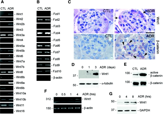
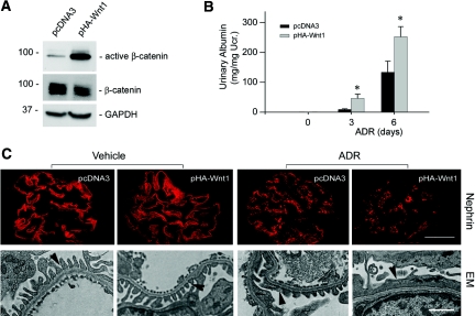
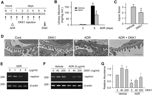
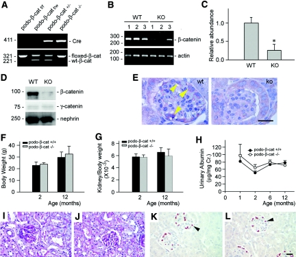
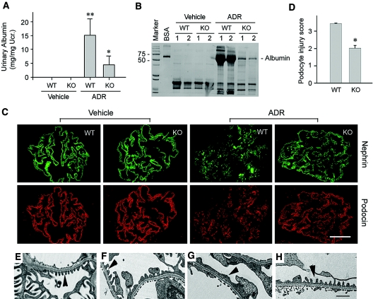
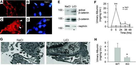
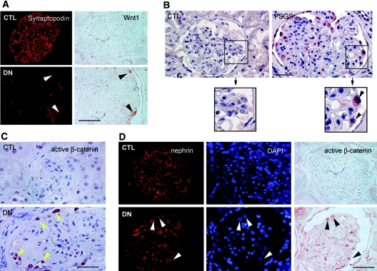
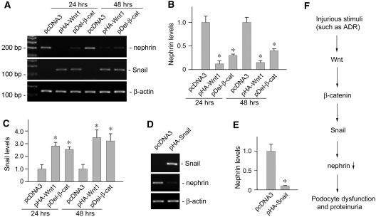
Podocyte dysfunction, one of the major causes of proteinuria, leads to glomerulosclerosis and end stage renal disease, but its underlying mechanism remains poorly understood. Here we show that Wnt/beta-catenin signaling plays a critical role in podocyte injury and proteinuria. Treatment with adriamycin induced Wnt and activated beta-catenin in mouse podocytes. Overexpression of Wnt1 in vivo activated glomerular beta-catenin and aggravated albuminuria and adriamycin-induced suppression of nephrin expression, whereas blockade of Wnt signaling with Dickkopf-1 ameliorated podocyte lesions. Podocyte-specific knockout of beta-catenin protected against development of albuminuria after injury. Moreover, pharmacologic activation of beta-catenin induced albuminuria in wild-type mice but not in beta-catenin-knockout littermates. In human proteinuric kidney diseases such as diabetic nephropathy and focal segmental glomerulosclerosis, we observed upregulation of Wnt1 and active beta-catenin in podocytes. Ectopic expression of either Wnt1 or stabilized beta-catenin in vitro induced the transcription factor Snail and suppressed nephrin expression, leading to podocyte dysfunction. These results suggest that targeting hyperactive Wnt/beta-catenin signaling may represent a novel therapeutic strategy for proteinuric kidney diseases.


Figures

Figure 1.
Figure 1.
Wnt/β-catenin signaling is activated in podocytes after injury in vivo and in vitro. (A and B) RT-PCR demonstrates an altered expression of various (A) Wnt and (B) Frizzled receptor (Fzd) mRNA in the glomeruli isolated from mice at 1 d after ADR injection. (C) Immunohistochemical staining shows Wnt1 and β-catenin induction in podocytes after ADR injury. Arrows indicate the Wnt1-positive cells. Arrowheads show the cytoplasmic and nuclear staining of β-catenin. Scale bar, 30 μm. (D) Western blot demonstrates an induction of Wnt1 protein in the glomeruli isolated from mice at different time points as indicated after ADR injection. (E) Activation of β-catenin signaling in the isolated glomeruli after ADR injection. Glomerular lysates were immunoblotted with antibodies against active and total β-catenin, respectively. (F and G) RT-PCR and Western blot demonstrate upregulation of Wnt1 (F) mRNA and (G) protein expression in podocytes after injury in vitro. Mouse podocytes were treated with ADR (10 μg/ml) for various periods of time as indicated.
Figure 2.
Figure 2.
Exogenous Wnt1 aggravates podocytes dysfunction and albuminuria in vivo. BALB/c mice were administrated with either Wnt1 expression vector (pHA-Wnt1) or control plasmid (pcDNA3) through a hydrodynamics-based tail vein injection. (A) Wnt1 gene delivery induces active β-catenin accumulation in the glomeruli. Glomerular lysates from the mice injected with either pcDNA3 or pHA-Wnt1 expression vector were immunoblotted with antibodies against active β-catenin, total β-catenin, and glyceraldehyde 3-phosphate dehydrogenase, respectively. (B) Exogenous Wnt1 exacerbates albuminuria in mice after ADR injection. *P < 0.05 versus pcDNA3 controls (n = 6). (C) Representative micrographs showing nephrin staining and ultrastructure of podocyte foot processes in different groups as indicated. Immunofluorescence staining shows nephrin protein expression and distribution in different groups. Scale bar = 30 μm. Podocyte foot processes integrity was revealed by EM. Arrowheads indicate secondary foot process and SD. Scale bar, 1 μm.
Figure 3.
Figure 3.
Blockade of Wnt signaling attenuates podocyte injury and proteinuria. (A) Experimental design showing the strategy of recombinant DKK1 protein and ADR injections in mice. (B and C) DKK1 reduces (B) ADR-induced albuminuria and (C) podocyte injury (as defined by nephrin loss and altered distribution) in mice. *P < 0.05 versus vehicle group (n = 8). (D) Representative micrographs showing podocyte foot processes in different groups as indicated. Arrowheads indicate secondary foot process and SD. Scale bar, 1 μm. (E through G) DKK1 restores nephrin expression after ADR treatment in cultured mouse podocytes. (E) ADR suppressed nephrin expression in a dose-dependent manner in podocytes. (F and G) DKK1 restored nephrin expression after ADR treatment. (F) Representative RT-PCR result and (G) relative abundances of nephrin mRNA (with value in control group = 1.0) are shown. *P < 0.05 versus controls; †P < 0.05 versus the group without DKK1 treatment (n = 3).
Figure 4.
Figure 4.
Mice with podocyte-specific deletion of β-catenin are healthy. (A) Genotyping of the mice by PCR analysis of genomic DNA. (B and C) RT-PCR analysis shows the glomerular β-catenin mRNA abundance in podo-β-cat−/− mice and their control littermates. WT, wild-type mice; KO, podo-β-cat−/− mice. Numbers (1, 2, and 3) denote each individual animal in a given group. *P < 0.05 versus WT (n = 3). (D) Western blot analysis of glomerular β-catenin protein in podo-β-cat−/− mice. Glomerular lysates were prepared from WT or KO mice and immunoblotted with antibodies against β-catenin, γ-catenin, and nephrin, respectively. (E) Immunohistochemical staining shows loss of β-catenin in glomerular podocytes. Arrowhead indicates positive staining for β-catenin in the glomeruli of WT mice at 1 d after ADR injection. No β-catenin staining was observed in the glomeruli of KO mice at the same conditions. (F through L) Mice with podocyte-specific ablation of β-catenin are phenotypically normal. There was little difference in (F) body weight, (G) kidney weight index, and (H) urinary albumin between KO mice and control littermates at different time points (n = 4 to 6). Kidney histology as shown by (I and J) periodic-acid-Schiff staining and (K and L) podocyte by WT-1 staining are normal in (J and L) KO mice, compared with (I and K) control littermates.
Figure 5.
Figure 5.
Podocyte-specific ablation of β-catenin protects against albuminuria and podocyte injury induced by ADR in mice. (A) Urinary albumin concentration in WT and KO mice at 3 d after ADR injection. **P < 0.01 versus vehicle control, *P < 0.05 versus WT mice after ADR injection (n = 4 to 9). (B) Representative SDS-PAGE shows the urine proteins in different groups of mice as indicated. Numbers (1 and 2) denote each individual animal in a given group. (C and D) Immunofluorescence staining demonstrates the abundance and distribution pattern of nephrin and podocin in different groups as indicated. (C) Representative micrographs and (D) semiquantitative determination of podocyte injury (as defined by nephrin loss and altered distribution) are given. *P < 0.05 versus WT group (n = 4 to 9). (E through H) EM shows the alterations of podocyte foot processes after ADR injection in WT and KO mice. (E) WT control, (F) KO control, (G) WT after ADR, and (H) KO after ADR. (G) Foot process effacement is evident in WT podocytes after ADR. Scale bar, 1 μm.
Figure 6.
Figure 6.
Activation of β-catenin signaling induces podocyte dysfunction and albuminuria in vivo. (A through D) Incubation with LiCl (30 mM) induces β-catenin activation and nuclear translocation in cultured podocytes. (A and B) NaCl control, (C and D) LiCl (30 mM), (A and C) β-catenin staining, and (B and D) DAPI. Arrowheads indicate nuclear staining of β-catenin after LiCl treatment. (E) Activation of β-catenin signaling in the glomeruli by LiCl in vivo. Glomerular lysates from the mice injected with either LiCl or control NaCl were immunoblotted with antibodies against active β-catenin, total β-catenin, and nephrin, respectively. (F) Activation of β-catenin by LiCl causes heavy, albeit transient, albuminuria in mice. **P < 0.01 versus NaCl control (n = 4 to 10). (G) EM shows podocyte foot process effacement in mice after LiCl injection. (H) Podocyte-specific ablation of β-catenin protects mice from development of albuminuria after LiCl injection. *P < 0.05 versus WT controls (n = 7).
Figure 7.
Figure 7.
Wnt/β-catenin signaling is activated in human proteinuric nephropathies. Human kidney specimens were immunostained with specific antibodies against Wnt1, synaptopodin, nephrin, and active β-catenin, respectively. (A) Representative micrographs of double staining for Wnt1 and synaptopodin in human kidney biopsy from patients with DN. Arrowheads indicate colocalization of Wnt1 and synaptopodin. Scale bar, 40 μm. (B) Representative micrographs demonstrate Wnt1 induction in the glomerular cells in human kidney biopsy from the patients with FSGS. Arrowheads within the boxed area indicate Wnt1-positive cells. Scale bar, 40 μm. (C) Representative micrographs show active β-catenin staining in the nuclei of glomerular cells in human kidney biopsy from patients with DN. Arrowheads (yellow) indicate active β-catenin-positive nuclei. Scale bar, 40 μm. (D) Representative micrographs show the nuclear staining of active β-catenin in podocytes in human kidney biopsy from the patients with DN. Arrowheads indicate the nephrin-positive podocytes with nuclear staining for active β-catenin. Scale bar, 40 μm. CTL, control.
Figure 8.
Figure 8.
Wnt/β-catenin signaling induces Snail expression and suppresses nephrin in podocytes. (A through C) Wnt1 and β-catenin are sufficient for triggering nephrin suppression and Snail induction in podocytes. Mouse podocytes were transiently transfected with Wnt1 expression vector (pHA-Wnt1), expression plasmid for the stabilized β-catenin with N-terminal deletion (pDel-β-cat), or empty vector pcDNA3, respectively, followed by analyzing nephrin and Snail expression. Representative results on (A) nephrin and Snail mRNA expression and quantitative data on relative (B) nephrin or (C) Snail levels (with value in pcDNA3 control group = 1.0) are presented. *P < 0.05 versus controls (n = 3). (D and E) Snail is sufficient for suppressing nephrin expression in podocytes. Mouse podocytes were transiently transfected with pHA-Snail or pcDNA3 plasmids, followed by analyzing nephrin expression. (D) Representative RT-PCR results and (E) quantitative determination of relative nephrin mRNA levels (with the value in the pcDNA3 control group = 1.0) are presented. *P < 0.05 versus controls (n = 4). (F) Simplified diagram depicts the molecular pathway by which Wnt/β-catenin signaling mediates podocyte dysfunction and proteinuria.

Comment in

References

    1. Coresh J, Selvin E, Stevens LA, Manzi J, Kusek JW, Eggers P, Van Lente F, Levey AS:Prevalence of chronic kidney disease in the United States. JAMA 298: 2038–2047, 2007 - PubMed
    1. Eknoyan G, Lameire N, Barsoum R, Eckardt KU, Levin A, Levin N, Locatelli F, MacLeod A, Vanholder R, Walker R, Wang H:The burden of kidney disease: Improving global outcomes. Kidney Int 66: 1310–1314, 2004 - PubMed
    1. Wiggins RC:The spectrum of podocytopathies: A unifying view of glomerular diseases. Kidney Int 71: 1205–1214, 2007 - PubMed
    1. Shankland SJ:The podocyte's response to injury: Role in proteinuria and glomerulosclerosis. Kidney Int 69: 2131–2147, 2006 - PubMed
    1. Kim JM, Wu H, Green G, Winkler CA, Kopp JB, Miner JH, Unanue ER, Shaw AS:CD2-associated protein haploinsufficiency is linked to glomerular disease susceptibility. Science 300: 1298–1300, 2003 - PubMed

Publication types

MeSH terms