. 2009 Aug 4;106(31):12706-11.
doi: 10.1073/pnas.0905046106. Epub 2009 Jul 23.

Triplex structures in an RNA pseudoknot enhance mechanical stability and increase efficiency of -1 ribosomal frameshifting

Affiliations

Triplex structures in an RNA pseudoknot enhance mechanical stability and increase efficiency of -1 ribosomal frameshifting

Gang Chen et al. Proc Natl Acad Sci U S A. .

Abstract

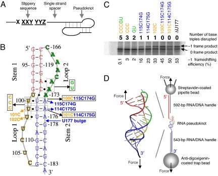
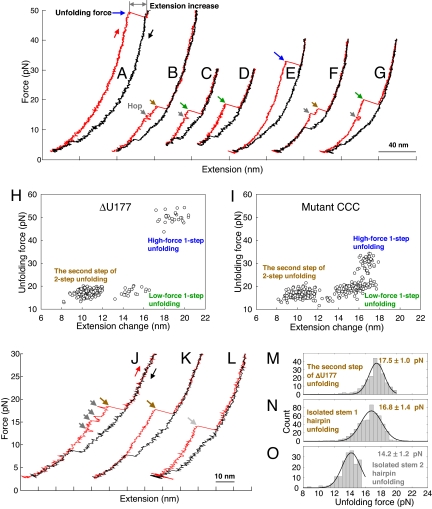
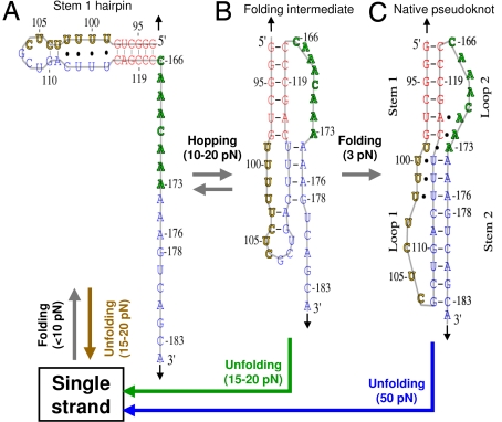
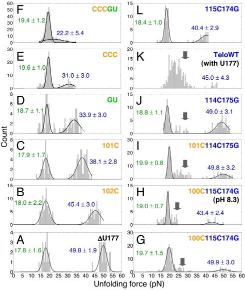
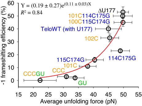
Many viruses use programmed -1 ribosomal frameshifting to express defined ratios of structural and enzymatic proteins. Pseudoknot structures in messenger RNAs stimulate frameshifting in upstream slippery sequences. The detailed molecular determinants of pseudoknot mechanical stability and frameshifting efficiency are not well understood. Here we use single-molecule unfolding studies by optical tweezers, and frameshifting assays to elucidate how mechanical stability of a pseudoknot and its frameshifting efficiency are regulated by tertiary stem-loop interactions. Mechanical unfolding of a model pseudoknot and mutants designed to dissect specific interactions reveals that mechanical stability depends strongly on triplex structures formed by stem-loop interactions. Combining single-molecule and mutational studies facilitates the identification of pseudoknot folding intermediates. Average unfolding forces of the pseudoknot and mutants ranging from 50 to 22 picoNewtons correlated with frameshifting efficiencies ranging from 53% to 0%. Formation of major-groove and minor-groove triplex structures enhances pseudoknot stem stability and torsional resistance, and may thereby stimulate frameshifting. Better understanding of the molecular determinants of frameshifting efficiency may facilitate the development of anti-virus therapeutics targeting frameshifting.


Conflict of interest statement

The authors declare no conflict of interest.

Figures

Fig. 1.
Fig. 1.
Minus-one ribosomal frameshifting (FS), pseudoknots, and experimental design. (A) Three components for programmed FS. (B) Pseudoknots used for bulk FS assay and single-molecule studies. All of the mutants were made based on ΔU177. The secondary structure of pseudoknot ΔU177 is also shown in Fig. 3C. In mutant CCCGU, all 5 base triples formed between stem 1 (shown in red) and loop 2 (shown in green) and between stem 2 (shown in blue) and loop 1 (shown in yellow) are disrupted. In TeloWT, the single nucleotide bulge U177 in stem 2 is added. (C) A typical SDS/PAGE result for the bulk FS assay. Shown on top and below the gel are the numbers of base triples disrupted and values of FS efficiency, respectively. (D) Experimental setup for mechanical (un)folding of RNA pseudoknots using optical tweezers. Pseudoknot ΔU177 is flanked by RNA/DNA hybrid handles, which are in turn attached to pipette bead and trap bead. The directions of applied force are shown with black arrows. Drawing is not to scale.
Fig. 2.
Fig. 2.
Representative force-extension curves and summary of mechanical unfolding results. Red and black curves are for pulling and relaxing, respectively, with waiting time at approximately 3 pN for 0 or 10 s. (A–D) Representative force-extension curves of ΔU177. (E–G) Representative force-extension curves of mutant CCC (with 3 major-groove U·A-U base triples disrupted, see Fig. 1B). (H) Summary of unfolding force-versus-extension change of ΔU177. (I) Summary of unfolding force-versus-extension change of mutant CCC. (J) A force-extension curve of ΔU177 folding intermediate structure with low-force 2-step unfolding. (K) A force-extension curve of isolated stem 1 hairpin derived from ΔU177 (see Fig. S4 A). (L) A force-extension curve of isolated stem 2 hairpin derived from ΔU177. (M) Unfolding force histogram of the second step of low-force 2-step unfolding of the folding intermediate structure of pseudoknot ΔU177. (N) Unfolding force histogram of isolated stem 1 hairpin derived from ΔU177 (see Fig. S4 B). (O) Unfolding force histogram of isolated stem 2 hairpin derived from ΔU177.
Fig. 3.
Fig. 3.
Possible folding intermediate structures of pseudoknot ΔU177. The positions and directions of applied force are indicated with black arrows. The unfolding transitions are indicated with the same colors as those in Fig. 2. (A) Stem 1 hairpin. In a force-ramp experiment, stem 1 hairpin typically folds from single strand when force is below 10 pN. Stem 1 hairpin unfolds to single strand at 15–20 pN. (B) A folding intermediate with stem 2 partially formed. In a force-ramp experiment, the folding intermediate rapidly forms after stem 1 hairpin is formed when force is below 10 pN. Upon increasing force to between 10 and 20 pN, the folding intermediate is in equilibrium with stem 1 hairpin and hopping between the 2 structures were observed. The folding intermediate may unfold apparently in 1 step or 2 steps at 15–20 pN. (C) Native pseudoknot. Folding transition from the folding intermediate to native pseudoknot is slow even at 3 pN as indicated by subsequent unfolding trajectories (see Fig. 2). Native pseudoknot unfolds to single strand in 1 step at approximately 50 pN.
Fig. 4.
Fig. 4.
Unfolding force histograms for 1-step unfolding reactions. Values shown are peak values of Gaussian distributions with standard deviations. Mutations have significant effect on the native pseudoknots (higher unfolding forces, shown in blue) but not the folding intermediate structures (lower unfolding forces, shown in green). With the entire 5 base triples disrupted, mutant CCCGU still has 2 clusters of unfolding forces (F). Folding intermediate structures appear for pseudoknots with mutations in stem 2 (see arrows). Multiple folding intermediates may exist for TeloWT (K). All of the unfolding forces were measured at pH 7.3 except for mutant 100C115C174G, which was also measured at pH 8.3 (see H).
Fig. 5.
Fig. 5.
A single exponential function fits well (R2 = 0.84) the correlation between FS efficiency and average unfolding force. Error bars for unfolding force are standard deviations from multiple measurements. Error bars for FS efficiency are standard errors.

Similar articles

Cited by

References

    1. Atkins JF, Elseviers D, Gorini L. Low activity of β-galactosidase in frameshift mutants of Escherichia coli. Proc Natl Acad Sci USA. 1972;69:1192–1195. - PMC - PubMed
    1. Jacks T, Varmus HE. Expression of the Rous sarcoma virus pol gene by ribosomal frameshifting. Science. 1985;230:1237–1242. - PubMed
    1. Jacks T, et al. Characterization of ribosomal frameshifting in HIV-1 gag-pol expression. Nature. 1988;331:280–283. - PubMed
    1. Baril M, Dulude D, Gendron K, Lemay G, Brakier-Gingras L. Efficiency of a programmed –1 ribosomal frameshift in the different subtypes of the human immunodeficiency virus type 1 group M. RNA. 2003;9:1246–1253. - PMC - PubMed
    1. Plant EP, Dinman JD. The role of programmed -1 ribosomal frameshifting in coronavirus propagation. Front Biosci. 2008;13:4873–4881. - PMC - PubMed

Publication types

LinkOut - more resources