. 2009 Aug 11;106(32):13570-5.
doi: 10.1073/pnas.0905125106. Epub 2009 Jul 23.

Targeting neural precursors in the adult brain rescues injured dopamine neurons

Affiliations

Targeting neural precursors in the adult brain rescues injured dopamine neurons

Andreas Androutsellis-Theotokis et al. Proc Natl Acad Sci U S A. .

Abstract

In Parkinson's disease, multiple cell types in many brain regions are afflicted. As a consequence, a therapeutic strategy that activates a general neuroprotective response may be valuable. We have previously shown that Notch ligands support neural precursor cells in vitro and in vivo. Here we show that neural precursors express the angiopoietin receptor Tie2 and that injections of angiopoietin2 activate precursors in the adult brain. Signaling downstream of Tie2 and the Notch receptor regulate blood vessel formation. In the adult brain, angiopoietin2 and the Notch ligand Dll4 activate neural precursors with opposing effects on the density of blood vessels. A model of Parkinson's disease was used to show that angiopoietin2 and Dll4 rescue injured dopamine neurons with motor behavioral improvement. A combination of growth factors with little impact on the vasculature retains the ability to stimulate neural precursors and protect dopamine neurons. The cellular and pharmacological basis of the neuroprotective effects achieved by these single treatments merits further analysis.


Conflict of interest statement

The authors declare no conflicts of interest.

Figures
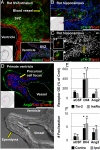
Fig. 1.
Fig. 1.
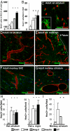
Neural precursors in the adult brain express the Tie2 receptor. (A) Phosphorylated Tie2 (pTie2) is expressed in the SVZ and blood vessels of the adult rat brain (Inset, nonphospho Tie2 expression in the SVZ). (B) pTie2 is co-expressed with Sox2 in the SGZ of the DG of the adult rat hippocampus (image from a CT-treated animal). (C) DAPI counterstain of the image in B and 2 single z planes, 4 μm apart (arrows point to the Sox2- positive /pTie2-positive nuclei). (D) A single Dll4 injection in the monkey striatum induces the formation of Tie2-rich foci at the surface of the lateral ventricle (image from a Dll4-treated monkey; below, DIC image of the same area). (E) Quantitation of Tie2 and insulin receptor subunit a signal of adult monkeys 10 days after single striatal injections of factors (data are ipsilateral OD signal from digital immunohistochemistry images, expressed as % contralateral striatum). (F) Quantitation of foci number in the ipsilateral and contralateral hemispheres of adult monkeys [data are total number of foci per coronal section of the SVZ (30 μm thickness)]. (Scale bars, 50 μm.)
Fig. 2.
Fig. 2.
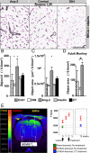
Angiogenic factors activate widespread neural precursors in vivo. (A) Single intracerebroventricular injections of soluble factors increase BrdU+ cell number in the SVZ 1 week following injection (numbers are % BSA sham injection controls) and (B) Hes3-positive cell number (numbers are % BSA sham controls). (C) Hes3-positive cells in the SVZ and striatum of control and treated animals co-express Sox2 (image from Dll4-treated striatum; inset, 3-D reconstruction from confocal z stacks). (D and E) Hes3-positive cells in the striatum and midbrain of adult rats are in contact with blood vessels (identified by RECA-1 expression; images are from CT-treated brains). (F and G) Hes3-positive cells in the SVZ and striatum of adult monkeys are in contact with blood vessels (identified by CD31 expression; images are from Ang2-treated brains). (H) Single intracerebroventricular injections of factors increase Hes3-positive cell number in the ipsilateral striatum and substantia nigra of adult rats (Hes3-positive cell numbers per field are shown). (I) Single intrastriatal MRI-guided convection delivery of factors increases the number of Hes3-positive cells in the ipsilateral striatum of adult monkeys (data are % BSA sham controls). (Scale bars: C, 50 μm; D–G, 100 μm.)
Fig. 3.
Fig. 3.
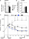
Combinations of pro- and anti-angiogenic factors maintain normal vascular density. (A) Thirteen weeks after a single intracerebroventricular injection of Ang2, there is an increase in blood vessel number and total vessel area in the striatum of adult rats; Dll4 induces a reduction in vessel number. (B and C) Quantitation of vascular density (“object number”) and total vessel surface area (“object area”) by pattern recognition software in striatal sections stained for RECA-1. (D) Quantitation of total vessel surface area (“object area”) in striatal sections from adult monkeys (10 days after a single intrastriatal injection of factors), stained for CD31. (E) CT treatment induces the normalization of T1 MRI signal following 6-OHDA lesion relative to sham-treated BSA controls [red bars, 6-OHDA; yellow bar, saline control (no 6-OHDA lesion); blue bars, 6-OHDA lesion followed by CT injections, 2 weeks later]. (Scale bar, 100 μm.)
Fig. 4.
Fig. 4.
Injured dopamine neurons are protected from death by single treatments with angiogenic factors. (A) Single intracerebroventricular injections of Dll4, Ang2, and CT result in an increase in TH+ signal in the lesioned rat striatum (data are expressed as % ipsilateral versus contralateral TH optical density; contralateral TH signal was similar in all treatment groups). (B) When administered 2 weeks after a 6-OHDA lesion, the same treatments also rescue TH+ cell bodies in the substantia nigra (assessed 13 weeks after treatment). Dopamine neurons were retrogradely labeled by fluorogold injections in the striatum, 1 week before euthanization; data are numbers of Fluorogold+/TH+ cell bodies in the substantia nigra as a % control. (C) Single intracerebroventricular injections of various treatments 2 weeks after 6-OHDA lesion in adult rats promote long-lasting behavioral recovery assessed by amphetamine-induced rotometry.

References

    1. Kim JH, et al. Dopamine neurons derived from embryonic stem cells function in an animal model of Parkinson's disease. Nature. 2002;418:50–56. - PubMed
    1. Perrier AL, et al. Derivation of midbrain dopamine neurons from human embryonic stem cells. Proc Natl Acad Sci USA. 2004;101:12543–12548. - PMC - PubMed
    1. Roy NS, et al. Functional engraftment of human ES cell-derived dopaminergic neurons enriched by coculture with telomerase-immortalized midbrain astrocytes. Nat Med. 2006;12:1259–1268. - PubMed
    1. Freed CR, et al. Transplantation of embryonic dopamine neurons for severe Parkinson's disease. N Engl J Med. 2001;344:710–719. - PubMed
    1. Hagell P, et al. Dyskinesias following neural transplantation in Parkinson's disease. Nat Neurosci. 2002;5:627–628. - PubMed

Publication types

MeSH terms