

Connexin 43 as a signaling platform for increasing the volume and spatial distribution of regenerated tissue
- PMID: 19628695
- PMCID: PMC2726403
- DOI: 10.1073/pnas.0902622106
Connexin 43 as a signaling platform for increasing the volume and spatial distribution of regenerated tissue
Abstract
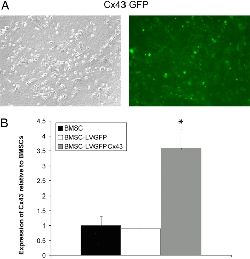
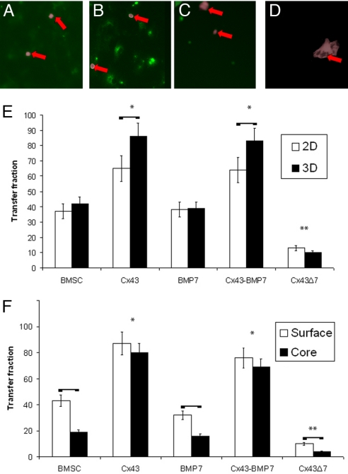
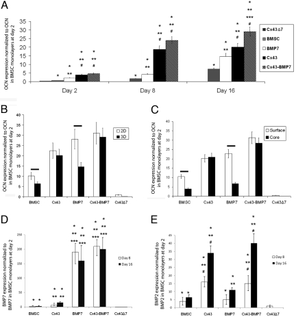
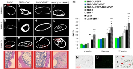
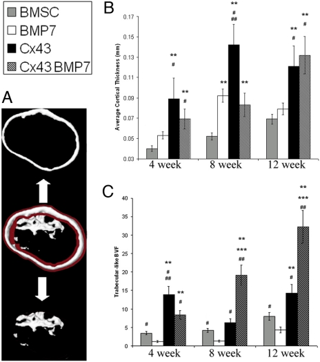
Gap junction intercellular communication (GJIC) is ubiquitous in the majority of vertebrate cells and is required for the proper development of most tissues. The loss of gap junction-mediated cell-to-cell communication leads to compromised development in many tissues and organs. Because cells constantly interact through gap junctions to coordinate tissue functions and homeostasis, we hypothesized that increasing cell-to-cell communication, via genetically engineering cells to overexpress gap junction proteins, could enhance cell differentiation in the interior regions of 3D tissue equivalents, thereby increasing the ability to regenerate larger and more uniform volumes of tissue. To test this hypothesis, we used bone as a model tissue because of the difficulty in achieving spatially uniform bone regeneration in 3D. In bone marrow stromal cells (BMSC), GJIC and osteogenic differentiation were compromised in 3D cultures relative to 2D monolayers and in the core of 3D cultures relative to the surface. Overexpression of connexin 43 (Cx43) via transduction of BMSCs with a lentivirus overcame this problem, enhancing both the magnitude and spatial distribution of GJIC and osteogenic differentiation markers throughout 3D constructs. Transplantation of cells overexpressing Cx43 resulted in an increased volume fraction and spatial uniformity of bone in vivo, relative to nontransduced BMSCs. Increased GJIC also enhanced the effect of a potent osteoinductive agent (BMP-7), suggesting a synergism between the soluble factor and GJIC. These findings present a platform to improve cell-to-cell communication in 3D and to achieve uniformly distributed tissue regeneration in 3D.
Conflict of interest statement
The authors declare no conflict of interest.
Figures
Similar articles
-
Cell communication and tissue engineering.Commun Integr Biol. 2010 Jan;3(1):53-6. doi: 10.4161/cib.3.1.9863. Commun Integr Biol. 2010. PMID: 20539784 Free PMC article.
-
Connexin43 intercellular communication drives the early differentiation of human bone marrow stromal cells into osteoblasts.J Cell Physiol. 2018 Feb;233(2):946-957. doi: 10.1002/jcp.25938. Epub 2017 May 23. J Cell Physiol. 2018. PMID: 28369869
-
The role of the micro-pattern and nano-topography of hydroxyapatite bioceramics on stimulating osteogenic differentiation of mesenchymal stem cells.Acta Biomater. 2018 Jun;73:509-521. doi: 10.1016/j.actbio.2018.04.030. Epub 2018 Apr 18. Acta Biomater. 2018. PMID: 29678674
-
Gap junctions and hemichannels in signal transmission, function and development of bone.Biochim Biophys Acta. 2012 Aug;1818(8):1909-18. doi: 10.1016/j.bbamem.2011.09.018. Epub 2011 Sep 22. Biochim Biophys Acta. 2012. PMID: 21963408 Free PMC article. Review.
-
Gap junction intercellular communication: a review of a potential platform to modulate craniofacial tissue engineering.J Biomed Mater Res B Appl Biomater. 2009 Feb;88(2):509-18. doi: 10.1002/jbm.b.31127. J Biomed Mater Res B Appl Biomater. 2009. PMID: 18481782 Free PMC article. Review.
Cited by
-
Mammalian genes induce partially reprogrammed pluripotent stem cells in non-mammalian vertebrate and invertebrate species.Elife. 2013 Sep 3;2:e00036. doi: 10.7554/eLife.00036. Elife. 2013. PMID: 24015354 Free PMC article.
-
ERK acts in parallel to PKCδ to mediate the connexin43-dependent potentiation of Runx2 activity by FGF2 in MC3T3 osteoblasts.Am J Physiol Cell Physiol. 2012 Apr 1;302(7):C1035-44. doi: 10.1152/ajpcell.00262.2011. Epub 2012 Jan 25. Am J Physiol Cell Physiol. 2012. PMID: 22277757 Free PMC article.
-
Quercetin stimulates osteogenic differentiation of bone marrow stromal cells through miRNA-206/connexin 43 pathway.Am J Transl Res. 2020 May 15;12(5):2062-2070. eCollection 2020. Am J Transl Res. 2020. PMID: 32509200 Free PMC article.
-
Engineering the matrix microenvironment for cell delivery and engraftment for tissue repair.Curr Opin Biotechnol. 2013 Oct;24(5):864-71. doi: 10.1016/j.copbio.2013.04.005. Epub 2013 May 4. Curr Opin Biotechnol. 2013. PMID: 23647972 Free PMC article. Review.
-
Low frequency‑pulsed electromagnetic fields promote osteogenic differentiation of bone marrow‑derived mesenchymal stem cells by regulating connexin 43 expression.Exp Ther Med. 2024 Oct 1;28(6):446. doi: 10.3892/etm.2024.12736. eCollection 2024 Dec. Exp Ther Med. 2024. PMID: 39386938 Free PMC article.
References
-
- Jorgensen NR, et al. Activation of L-type calcium channels is required for Gap junction-mediated intercellular calcium signaling in osteoblastic cells. J Biol Chem. 2003;278:4082–4086. - PubMed
-
- Jongsma HJ, Wilders R. Gap junctions in cardiovascular disease. Circ Res. 2000;86:1193–1197. - PubMed
-
- Flenniken AM, et al. A Gja1 missense mutation in a mouse model of oculodentodigital dysplasia. Development. 2005;132:4375–4386. - PubMed
-
- King TJ, Bertram JS. Connexins as targets for cancer chemoprevention and chemotherapy. Biochim Biophys Acta. 2005;1719:146–160. - PubMed
Publication types
MeSH terms
Substances
Grants and funding
LinkOut - more resources
Full Text Sources
Other Literature Sources
Miscellaneous
