. 2009 Aug 11;106(32):13517-22.
doi: 10.1073/pnas.0906413106. Epub 2009 Jul 23.

Translation and replication of hepatitis C virus genomic RNA depends on ancient cellular proteins that control mRNA fates

Affiliations

Translation and replication of hepatitis C virus genomic RNA depends on ancient cellular proteins that control mRNA fates

Nicoletta Scheller et al. Proc Natl Acad Sci U S A. .

Abstract

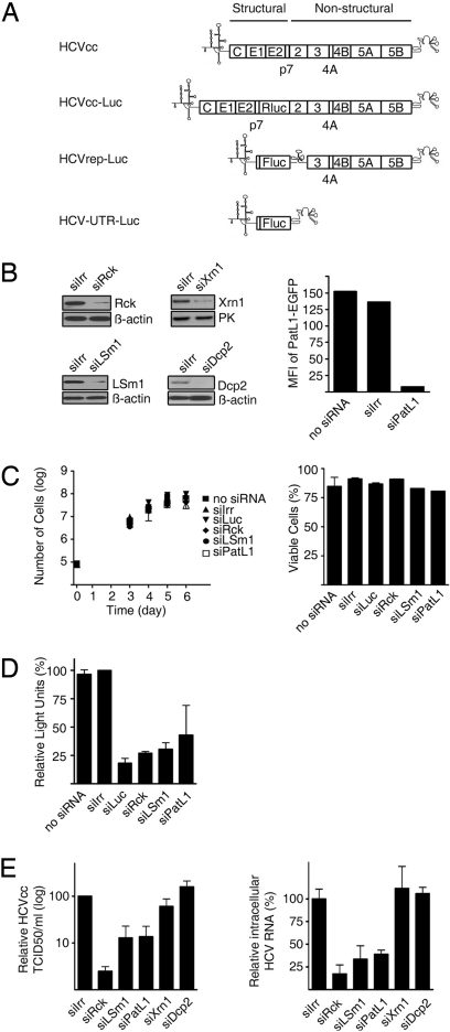
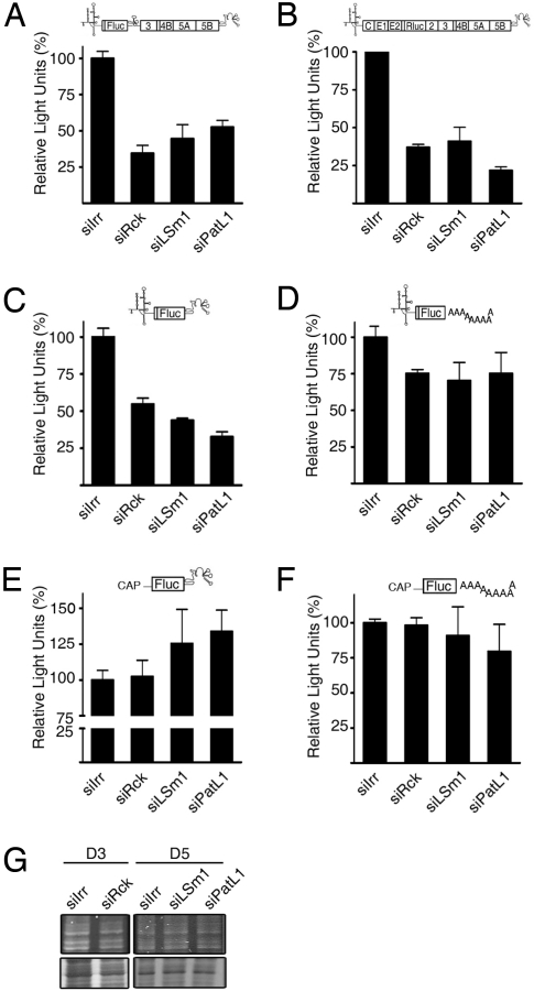
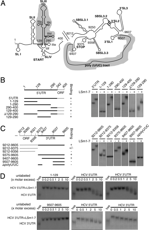
Inevitably, viruses depend on host factors for their multiplication. Here, we show that hepatitis C virus (HCV) RNA translation and replication depends on Rck/p54, LSm1, and PatL1, which regulate the fate of cellular mRNAs from translation to degradation in the 5'-3'-deadenylation-dependent mRNA decay pathway. The requirement of these proteins for efficient HCV RNA translation was linked to the 5' and 3' untranslated regions (UTRs) of the viral genome. Furthermore, LSm1-7 complexes specifically interacted with essential cis-acting HCV RNA elements located in the UTRs. These results bridge HCV life cycle requirements and highly conserved host proteins of cellular mRNA decay. The previously described role of these proteins in the replication of 2 other positive-strand RNA viruses, the plant brome mosaic virus and the bacteriophage Qss, pinpoint a weak spot that may be exploited to generate broad-spectrum antiviral drugs.


Conflict of interest statement

The authors declare no conflict of interest.

Figures

Fig. 1.
Fig. 1.
Depletion of Rck/p54, LSm1 or PatL1 in hepatoma cell lines impairs HCV replication. (A) Schematic representations of the genomes of HCVcc, HCV Replicons and derivatives used in this study. (B) Huh7-Lunet cells were transfected with siRNA targeting Rck/p54, LSm1, PatL1, Xrn1, Dcp2, or a nontargeting siRNA (siIrr). Immunoblot analyses of Rck/p54, LSm1, Xrn1, Dcp2, β-actin, or pyruvate kinase levels are shown. Because no specific antibody is available for PatL1, to test PatL1 silencing, PatL1-EGFP expression plasmid and siRNAs were cotransfected and fluorescence was analyzed 1 day later by flow cytometry. Values are expressed in mean fluorescence intensity (MFI) (bar graph). Similar silencing results were obtained for Huh7.5 cells. (C) Cell growth of siRNA-transfected cells was followed for 6 days by counting the total number of cells (mean ± SEM; n = 3) (Left). The percentage of viable silenced cells at the day of maximum silencing was measured by propidium iodide staining (mean ± SEM; n = 2) (Right). (D) Huh7-Lunet cells were coelectroporated with the HCVrep-Luc replicon and the siRNAs. The percentage of relative luciferase light units compared with siIrr-transfected cells is shown at the day of most efficient silencing (mean ± SEM; n = 2). (E) Three days after transfection of silenced Huh7.5 cells with HCVcc RNA, the HCVcc infectivity in the supernatant was titrated by a limited dilution assay (Left). The accumulation of intracellular HCVcc mRNA was analyzed by quantitative RT-PCR (Right). Both values were normalized to the amount of transfected RNA (mean ± SEM; n = 3) and are shown relative to siIrr-transfected cells.
Fig. 2.
Fig. 2.
Rck/p54, LSm1 and PatL1 silencing influences HCV RNA translation. Huh7-Lunet and Huh7.5 cells were transfected with siRNAs targeting Rck/p54, LSm1, PatL1, or a nontargeting siRNA, siIrr. The silenced cells were further transfected with (A) a nonreplicating bicistronic Luciferase replicon (HCVrep-Luc-GND), (B) a nonreplicating Luciferase-HCVcc (HCVcc-Luc-GNN), (C) a derivative (HCV-UTRs-Luc) containing the HCV 5′ and 3′UTRs from genotype 1b flanking the firefly luciferase ORF, (D) a derivative from HCV-UTRs-Luc in which the HCV 3′ UTR was exchanged by a poly(A) tail, (E) a derivative from HCV-UTRs-Luc in which the HCV 5′ UTR was exchanged by capped, nonviral 5′UTR, and (F) a derivative [CAP-Luc-Poly(A)] containing the 5‘capped, nonviral 5′UTR followed by the firefly luciferase ORF and a poly(A) tail. The luciferase activity was measured 4 h after transfection and normalized to the respective intracellular RNA levels measured by quantitative RT-PCR (mean ± SEM; n = 3). (G) To examine the influence of Rck/p54-, LSm1- and PatL1-silencing on the synthesis of cellular proteins, silenced cells were labeled with [35S]methionine for 30 min, separated on a denaturating polyacrylamide gel and visualized by autoradiography (Lower). Gels were coomassie-stained to visualize protein-loading (Upper).
Fig. 3.
Fig. 3.
Reconstituted LSm1–7 rings bind to specific HCV 5′ and 3′UTR regions. (A) Schematic representation of the secondary structures of the HCV 5′ and 3′ ends. Upstream of the 3′UTR, the NS5B coding sequence containing an RNA cruciform structure is shown. This structure includes the 5BSL3.2 loop. Its long-range interaction with the 3′SL3 loop in the 3′Xtail of the 3′UTR is essential for replication. Shadowed regions highlight the binding sites of the LSm1–7 rings. (B and C, Left) The constructs used in the electromobility shift assays are shown. The numbers refer to the nucleotide positions in the genome of the HCV Con1 strain. (B and C, Right) Radiolabeled, gel-purified RNA transcripts were incubated with the reconstituted LSm1–7 rings. After complex formation, products were separated on a nondenaturating polyacrylamide gel and visualized by autoradiography. (D) Labeled HCV 5′ and 3′UTR RNAs (HCV sequences 1–400 and 9,375–9,605, respectively) were incubated with reconstituted LSm1–7 rings in the presence of increasing amounts of unlabeled HCV 5′ and 3′UTR transcripts. As noncompeting controls, unlabeled RNAs negative for LSm1–7 ring binding (HCV sequences 1–129 and 9,507–9,605) were used. After complex formation, products were treated as in B and C.

Similar articles

Cited by

References

    1. Gamarnik AV, Andino R. Switch from translation to RNA replication in a positive-stranded RNA virus. Genes Dev. 1998;12:2293–2304. - PMC - PubMed
    1. Alves-Rodrigues I, Galao RP, Meyerhans A, Diez J. Saccharomyces cerevisiae: A useful model host to study fundamental biology of viral replication. Virus Res. 2006;120:49–56. - PMC - PubMed
    1. Diez J, Ishikawa M, Kaido M, Ahlquist P. Identification and characterization of a host protein required for efficient template selection in viral RNA replication. Proc Natl Acad Sci USA. 2000;97:3913–3918. - PMC - PubMed
    1. Mas A, et al. Host deadenylation-dependent mRNA decapping factors are required for a key step in brome mosaic virus RNA replication. J Virol. 2006;80:246–251. - PMC - PubMed
    1. Noueiry AO, et al. Yeast Lsm1p–7p/Pat1p deadenylation-dependent mRNA-decapping factors are required for brome mosaic virus genomic RNA translation. Mol Cell Biol. 2003;23:4094–4106. - PMC - PubMed

Publication types

MeSH terms