

Minimal requirement for induction of natural cytotoxicity and intersection of activation signals by inhibitory receptors
- PMID: 19628705
- PMCID: PMC2756125
- DOI: 10.1182/blood-2009-01-201632
Minimal requirement for induction of natural cytotoxicity and intersection of activation signals by inhibitory receptors
Abstract
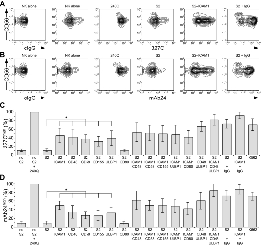
Natural killer (NK) cells provide innate control of infected and neoplastic cells. Multiple receptors have been implicated in natural cytotoxicity, but their individual contribution remains unclear. Here, we studied the activation of primary, resting human NK cells by Drosophila cells expressing ligands for receptors NKG2D, DNAM-1, 2B4, CD2, and LFA-1. Each receptor was capable of inducing inside-out signals for LFA-1, promoting adhesion, but none induced degranulation. Rather, release of cytolytic granules required synergistic activation through coengagement of receptors, shown here for NKG2D and 2B4. Although engagement of NKG2D and 2B4 was not sufficient for strong target cell lysis, collective engagement of LFA-1, NKG2D, and 2B4 defined a minimal requirement for natural cytotoxicity. Remarkably, inside-out signaling induced by each one of these receptors, including LFA-1, was inhibited by receptor CD94/NKG2A binding to HLA-E. Strong inside-out signals induced by the combination of NKG2D and 2B4 or by CD16 could overcome CD94/NKG2A inhibition. In contrast, degranulation induced by these receptors was still subject to inhibition by CD94/NKG2A. These results reveal multiple layers in the activation pathway for natural cytotoxicity and that steps as distinct as inside-out signaling to LFA-1 and signals for granule release are sensitive to inhibition by CD94/NKG2A.
Figures
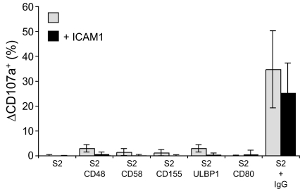
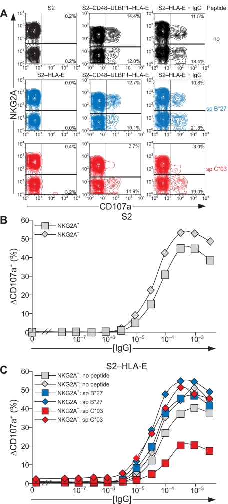
 represents target cells as indicated, whereas ■ represents target cells coexpressing ICAM-1 in addition to the other ligands, as indicated. Bars denote SD.
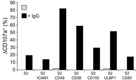
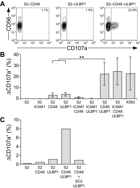
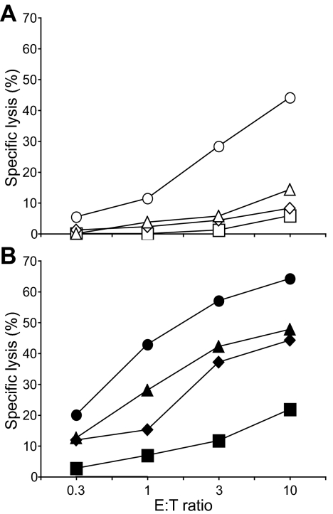
 ), S2-CD48-ULBP1 (△), or S2–ICAM-1–CD48–ULBP1 (○) cells, or (B) S2 cells as in panel A preincubated with a rabbit serum raised against S2 cells (■, ♦, ▲, ●). Cells were incubated for 3 hours at 37°C. Specific lysis of S2 cells was calculated from the percentage of propidium iodide–positive S2 cells in duplicate samples, as determined by flow cytometry. The experiment is representative of 3 or more independent experiments.
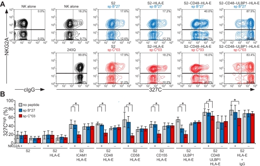
References
Publication types
MeSH terms
Substances
Grants and funding
LinkOut - more resources
Full Text Sources
Other Literature Sources
Research Materials
