The fibrotic phenotype induced by IGFBP-5 is regulated by MAPK activation and egr-1-dependent and -independent mechanisms
- PMID: 19628764
- PMCID: PMC2716960
- DOI: 10.2353/ajpath.2009.080991
The fibrotic phenotype induced by IGFBP-5 is regulated by MAPK activation and egr-1-dependent and -independent mechanisms
Abstract
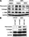
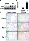
We have previously shown that insulin-like growth factor (IGF) binding protein- 5 (IGFBP-5) is overexpressed in lung fibrosis and induces the production of extracellular matrix components, such as collagen and fibronectin, both in vitro and in vivo. The exact mechanism by which IGFBP-5 exerts these novel fibrotic effects is unknown. We thus examined the signaling cascades that mediate IGFBP-5-induced fibrosis. We demonstrate for the first time that IGFBP-5 induction of extracellular matrix occurs independently of IGF-I, and results from IGFBP-5 activation of MAPK signaling, which facilitates the translocation of IGFBP-5 to the nucleus. We examined the effects of IGFBP-5 on early growth response (Egr)-1, a transcription factor that is central to growth factor-mediated fibrosis. Egr-1 was up-regulated by IGFBP-5 in a MAPK-dependent manner and bound to nuclear IGFBP-5. In fibroblasts from Egr-1 knockout mice, induction of fibronectin by IGFBP-5 was abolished. Expression of Egr-1 in these cells rescued the extracellular matrix-promoting effects of IGFBP-5. Moreover, IGFBP-5 induced cell migration in an Egr-1-dependent manner. Notably, Egr-1 levels, similar to IGFBP-5, were increased in vivo in lung tissues and in vitro in primary fibroblasts of patients with pulmonary idiopathic fibrosis. Taken together, our findings suggest that IGFBP-5 induces a fibrotic phenotype via the activation of MAPK signaling and the induction of nuclear Egr-1 that interacts with IGFBP-5 and promotes fibrotic gene transcription.
Figures


Similar articles
-
The pro-fibrotic factor IGFBP-5 induces lung fibroblast and mononuclear cell migration.Am J Respir Cell Mol Biol. 2009 Aug;41(2):179-88. doi: 10.1165/rcmb.2008-0211OC. Epub 2009 Jan 8. Am J Respir Cell Mol Biol. 2009. PMID: 19131643 Free PMC article.
-
NADPH oxidase-mediated induction of reactive oxygen species and extracellular matrix deposition by insulin-like growth factor binding protein-5.Am J Physiol Lung Cell Mol Physiol. 2019 Apr 1;316(4):L644-L655. doi: 10.1152/ajplung.00106.2018. Epub 2019 Feb 27. Am J Physiol Lung Cell Mol Physiol. 2019. PMID: 30810066 Free PMC article.
-
Insulin-like growth factor-binding protein-5 induces pulmonary fibrosis and triggers mononuclear cellular infiltration.Am J Pathol. 2006 Nov;169(5):1633-42. doi: 10.2353/ajpath.2006.060501. Am J Pathol. 2006. PMID: 17071587 Free PMC article.
-
Early growth response transcription factors: key mediators of fibrosis and novel targets for anti-fibrotic therapy.Matrix Biol. 2011 May;30(4):235-42. doi: 10.1016/j.matbio.2011.03.005. Epub 2011 Apr 13. Matrix Biol. 2011. PMID: 21511034 Free PMC article. Review.
-
Egr-1: new conductor for the tissue repair orchestra directs harmony (regeneration) or cacophony (fibrosis).J Pathol. 2013 Jan;229(2):286-97. doi: 10.1002/path.4131. Epub 2012 Dec 3. J Pathol. 2013. PMID: 23132749 Free PMC article. Review.
Cited by
-
Elucidating the cellular mechanism for E2-induced dermal fibrosis.Arthritis Res Ther. 2021 Feb 27;23(1):68. doi: 10.1186/s13075-021-02441-x. Arthritis Res Ther. 2021. PMID: 33640015 Free PMC article.
-
Insulin-like growth factor binding protein 5b of Trachinotus ovatus and its heparin-binding motif play a critical role in host antibacterial immune responses via NF-κB pathway.Front Immunol. 2023 Feb 14;14:1126843. doi: 10.3389/fimmu.2023.1126843. eCollection 2023. Front Immunol. 2023. PMID: 36865533 Free PMC article.
-
The role of cancer-associated myofibroblasts in intrahepatic cholangiocarcinoma.Nat Rev Gastroenterol Hepatol. 2011 Nov 29;9(1):44-54. doi: 10.1038/nrgastro.2011.222. Nat Rev Gastroenterol Hepatol. 2011. PMID: 22143274 Review.
-
Systems analysis of transcriptional data provides insights into muscle's biological response to botulinum toxin.Muscle Nerve. 2014 Nov;50(5):744-58. doi: 10.1002/mus.24211. Epub 2014 Mar 17. Muscle Nerve. 2014. PMID: 24536034 Free PMC article.
-
The Potential Use of Cannabis in Tissue Fibrosis.Front Cell Dev Biol. 2021 Oct 11;9:715380. doi: 10.3389/fcell.2021.715380. eCollection 2021. Front Cell Dev Biol. 2021. PMID: 34708034 Free PMC article. Review.
References
-
- Katzenstein AL, Myers JL. Idiopathic pulmonary fibrosis: clinical relevance of pathologic classification. Am J Respir Crit Care Med. 1998;157:1301–1315. - PubMed
-
- American Thoracic Society Idiopathic pulmonary fibrosis: diagnosis and treatment. International consensus statement. American Thoracic Society (ATS), and the European Respiratory Society (ERS). Am J Respir Crit Care Med. 2000;161:646–664. - PubMed
-
- Flaherty KR, Travis WD, Colby TV, Toews GB, Kazerooni EA, Gross BH, Jain A, Strawderman RL, Flint A, Lynch JP, Martinez FJ. Histopathologic variability in usual and nonspecific interstitial pneumonias. Am J Respir Crit Care Med. 2001;164:1722–1727. - PubMed
-
- Bjoraker JA, Ryu JH, Edwin MK, Myers JL, Tazelaar HD, Schroeder DR, Offord KP. Prognostic significance of histopathologic subsets in idiopathic pulmonary fibrosis. Am J Respir Crit Care Med. 1998;157:199–203. - PubMed
-
- Steen VD, Medsger TA. Severe organ involvement in systemic sclerosis with diffuse scleroderma. Arthritis Rheum. 2000;43:2437–2444. - PubMed
Publication types
MeSH terms
Substances
Grants and funding
LinkOut - more resources
Full Text Sources
Medical
Molecular Biology Databases

