. 2009 Oct 15;180(8):780-7.
doi: 10.1164/rccm.200810-1662OC. Epub 2009 Jul 23.

Evidence of dysfunction of endothelial progenitors in pulmonary arterial hypertension

Affiliations

Evidence of dysfunction of endothelial progenitors in pulmonary arterial hypertension

Mark Toshner et al. Am J Respir Crit Care Med. .

Abstract

Rationale: Severe pulmonary arterial hypertension (PAH) is characterized by the formation of plexiform lesions and concentric intimal fibrosis in small pulmonary arteries. The origin of cells contributing to these vascular lesions is uncertain. Endogenous endothelial progenitor cells are potential contributors to this process.

Objectives: To determine whether progenitors are involved in the pathobiology of PAH.

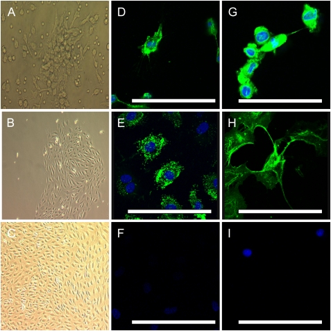
Methods: We performed immunohistochemistry to determine the expression of progenitor cell markers (CD133 and c-Kit) and the major homing signal pathway stromal cell-derived factor-1 and its chemokine receptor (CXCR4) in lung tissue from patients with idiopathic PAH, familial PAH, and PAH associated with congenital heart disease. Two separate flow cytometric methods were employed to determine peripheral blood circulating numbers of angiogenic progenitors. Late-outgrowth progenitor cells were expanded ex vivo from the peripheral blood of patients with mutations in the gene encoding bone morphogenetic protein receptor type II (BMPRII), and functional assays of migration, proliferation, and angiogenesis were undertaken. measurements and main results: There was a striking up-regulation of progenitor cell markers in remodeled arteries from all patients with PAH, specifically in plexiform lesions. These lesions also displayed increased stromal cell-derived factor-1 expression. Circulating angiogenic progenitor numbers in patients with PAH were increased compared with control subjects and functional studies of late-outgrowth progenitor cells from patients with PAH with BMPRII mutations revealed a hyperproliferative phenotype with impaired ability to form vascular networks.

Conclusions: These findings provide evidence of the involvement of progenitor cells in the vascular remodeling associated with PAH. Dysfunction of circulating progenitors in PAH may contribute to this process.


Figures

Figure 1.
Figure 1.
(AF) Representative photomicrographs of serial sections of lung tissue from control lung and a patient with familial pulmonary arterial hypertension (PAH); samples were immunostained for the endothelial marker CD31, and for CD133. Panels show staining in (A and B) a normal artery, (C and D) a concentric lesion, and (E and F) a plexiform lesion. (G) Semiquantitative analysis of CD133 expression in control arteries and in the vascular lesions of PAH. Box plots show median values, and upper and lower quartiles with standard error bars. Scale bars: 50 μm. ***P = 0.001.
Figure 2.
Figure 2.
Representative photomicrographs of confocal immunofluorescence staining of peripheral lung tissue in a patient with idiopathic pulmonary arterial hypertension (PAH): (AC) CD31 conjugated with fluorescein isothiocyanate (green) in (A) peripheral lung and (B and C) plexiform lesions. In the same sections (D) demonstrates CD133 in peripheral lung with isolated nonendothelial cells staining only in red (Texas red). (E) shows a plexiform lesion with CD133 staining the endothelial cell surface. (F) demonstrates a similar pattern of endothelial cell surface expression of c-Kit (Texas red). (GI) demonstrate that CD31 colocalizes with (H) CD133 and (I) c-Kit on the endothelial cell surface in both plexiform lesions when compared with nonplexiform peripheral lung tissue. TO-PRO-3 iodide counterstained for nuclear staining (blue). Scale bars: 50 μm.
Figure 3.
Figure 3.
Representative photomicrographs of peripheral lung tissue from (A and B) normal control lung and (CH) a patient with pulmonary arterial hypertension (PAH); samples were immunostained for CXCR4 and stromal cell–derived factor (SDF)-1. Minimal staining is seen in normal lung. (C and D) In PAH lung low-level staining was observed in concentric intimal lesions. (E and G) CXCR4 expression was generally increased in the lung parenchyma of patients with PAH, but was also present in the endothelium of plexiform lesions. (F and H) SDF-1 was less prevalent but showed clear staining of the endothelium of plexiform lesions. Scale bars: 50 μm.
Figure 4.
Figure 4.
Gating strategy for flow cytometry of CD133+CD34+VEFGR2+ cells by direct analysis of whole blood. A minimum of 200,000 events was recorded. (A) In a control subject a gate was drawn around the mononuclear cells. (B) Subsequently gates were drawn around the high-expressing distinct population of CD34+ cells, and (C) triple-positive events for CD34, CD133, and VEGFR2 were determined. (DF) Representative gates in a subject with idiopathic pulmonary arterial hypertension (IPAH). Events were included as positive only if exclusively triple positive on PRISM (Beckman Coulter analysis software). (G) Numbers of circulating CD133+CD34+VEGFR2+ cells per milliliter of blood in control subjects (n = 7), patients with IPAH (n = 7), and patients with PAH with bone morphogenetic protein receptor type II (BMPRII) mutations (n = 4). (H) Undifferentiated CD133+CD34+VEGFR2 circulating cells per milliliter of blood. Data are shown as means ± SEM (*P < 0.05).
Figure 5.
Figure 5.
Flow cytometric quantification of circulating putative endothelial progenitor cells (EPCs), using the purified CD133 fraction. Mononuclear cells were purified for CD133 antigen expression by magnetic cell sorting and double stained for CD133 and VEGFR2 (KDR). (A) Gating of EPCs after magnetic cell sorting. (B) Dead cells were excluded from analysis by their uptake of 7-amino-actinomycin D (-AAD). (C) Regions for fluorescence analysis were set with the negative control cells, and a maximum of 0.1% CD133+KDR+ cells was accepted in the negative control figure. (D) Cell preparation with 94.28% CD133-positive cells and a subset of 3.12% EPCs. (E) Quantification of circulating CD133+VEGFR2+ cells in the peripheral blood of control subjects (n = 11), patients with idiopathic pulmonary arterial hypertension (IPAH) (n = 10), and patients with secondary pulmonary arterial hypertension (SPAH, n = 13). Data are shown as means ± SEM (**P < 0.01).
Figure 6.
Figure 6.
Phase-contrast photomicrographs of cultured late-outgrowth endothelial progenitor cells (EPCs) showing (A) a colony-forming unit at 3 days, and a late-outgrowth colony at (B) 2 weeks and (C) 3 weeks. Confocal immunofluorescence images using conjugated fluorescein isothiocyanate (green) demonstrate that occasional cells were positive for (D) CD133 and that the majority of cells were positive for (E) von Willebrand factor, (G) CD34, and (H) CD146. Nuclear counterstaining was performed with 4′,6-diamidino-2-phenylindole (blue). (F and I) Isotype controls for anti-mouse and anti-rabbit secondary antibodies. Scale bars: 50 μm.
Figure 7.
Figure 7.
Phenotypic analysis of late-outgrowth endothelial progenitor cells (EPCs) from control subjects and patients with pulmonary arterial hypertension (PAH) associated with bone morphogenetic protein receptor type II (BMPRII) mutations. (A) Growth curves demonstrate that cells from patients with BMPRII-associated PAH are hyperproliferative, compared with those from control subjects, when grown in 10% fetal calf serum supplemented with additional growth factors. (B) Migration assays demonstrate that stromal cell–derived factor (SDF)-1 stimulated similar migration of cells from control subjects and subjects with BMPRII-associated PAH. Shaded columns, control subjects; solid columns, subjects with BMPRII mutation. HPF = high-power field. (C and D) Angiogenesis assays clearly demonstrate that (C) control EPCs form intact vascular networks, whereas (D) PAH BMPRII mutant EPCs are markedly deficient. (E) Semiquantitative analysis of vascular network formation. Experiments were performed in quadruplicate, n = 3 control and mutation cell lines. Data are shown as means ± SEM (*P < 0.05, ***P < 0.001).

References

    1. Voelkel NF, Cool C, Taraceviene-Stewart L, Geraci MW, Yeager M, Bull T, Kasper M, Tuder RM. Janus face of vascular endothelial growth factor: the obligatory survival factor for lung vascular endothelium controls precapillary artery remodeling in severe pulmonary hypertension. Crit Care Med 2002;30:S251–S256. - PubMed
    1. Wagenvoort CA, Wagenvoort N. Primary pulmonary hypertension: a pathologic study of the lung vessels in 156 clinically diagnosed cases. Circulation 1970;42:1163–1184.
    1. Lee SD, Shroyer KR, Markham NE, Cool CD, Voelkel NF, Tuder RM. Monoclonal endothelial cell proliferation is present in primary but not secondary pulmonary hypertension. J Clin Invest 1998;101:927–934. - PMC - PubMed
    1. Palevsky HI, Schloo BL, Pietra GG, Weber KT, Janicki JS, Rubin E, Fishman AP. Primary pulmonary hypertension: vascular structure, morphometry, and responsiveness to vasodilator agents. Circulation 1989;80:1207–1221. - PubMed
    1. Cool CD, Stewart JS, Werahera P, Miller GJ, Williams RL, Voelkel NF, Tuder RM. Three-dimensional reconstruction of pulmonary arteries in plexiform pulmonary hypertension using cell-specific markers: evidence for a dynamic and heterogeneous process of pulmonary endothelial cell growth. Am J Pathol 1999;155:411–419. - PMC - PubMed

Publication types

MeSH terms

Substances