. 2009 Aug 28;105(5):431-41.
doi: 10.1161/CIRCRESAHA.109.203083. Epub 2009 Jul 23.

Epicardium and myocardium separate from a common precursor pool by crosstalk between bone morphogenetic protein- and fibroblast growth factor-signaling pathways

Affiliations

Epicardium and myocardium separate from a common precursor pool by crosstalk between bone morphogenetic protein- and fibroblast growth factor-signaling pathways

Bram van Wijk et al. Circ Res. .

Abstract

Rationale: The epicardium contributes to the majority of nonmyocardial cells in the adult heart. Recent studies have reported that the epicardium is derived from Nkx2.5-positive progenitors and can differentiate into cardiomyocytes. Not much is known about the relation between the myocardial and epicardial lineage during development, whereas insights into these embryonic mechanisms could facilitate the design of future regenerative strategies.

Objective: Acquiring insight into the signaling pathways involved in the lineage separation leading to the differentiation of myocardial and (pro)epicardial cells at the inflow of the developing heart.

Methods and results: We made 3D reconstructions of Tbx18 gene expression patterns to give insight into the developing epicardium in relation to the developing myocardium. Next, using DiI tracing, we show that the (pro)epicardium separates from the same precursor pool as the inflow myocardium. In vitro, we show that this lineage separation is regulated by a crosstalk between bone morphogenetic protein (BMP) signaling and fibroblast growth factor (FGF) signaling. BMP signaling via Smad drives differentiation toward the myocardial lineage, which is inhibited by FGF signaling via mitogen-activated protein kinase kinase (Mek)1/2. Embryos exposed to recombinant FGF2 in vivo show enhanced epicardium formation, whereas a misbalance between FGF and BMP by Mek1/2 inhibition and BMP stimulation causes a developmental arrest of the epicardium and enhances myocardium formation at the inflow of the heart.

Conclusion: Our data show that FGF signaling via Mek1/2 is dominant over BMP signaling via Smad and is required to separate the epicardial lineage from precardiac mesoderm. Consequently, myocardial differentiation requires BMP signaling via Smad and inhibition of FGF signaling at the level of Mek1/2. These findings are of clinical interest for the development of regeneration-based therapies for heart disease.


Figures

Figure 1
Figure 1
Expression patterns of Tbx18 at the inflow of the heart. Tbx18 mRNA expression starts in the splanchnic mesoderm covering the vitelline veins at stage 11 (a and b). At stage 16, proepicardial and myocardial cells of the inflow are Tbx18-positive (c and d). One day later, stage 21, the epicardium and the myocardial sleeves covering the sinus horns are Tbx18 positive (e and f). Dashed lines indicate the position of the respective sections. lvv indicates left vitelline vein; rvv, right vitelline vein; nt, neural tube; fg, foregut; ift, inflow tract; oft, out flow tract; ra, right atrium; la, left atrium; v, ventricle; pe, proepicardium; rcv, right cardinal vein; lcv, left cardinal vein; vv, vitelline vein; li, liver; epi, epicardium.
Figure 2
Figure 2
Tracing the splanchnic mesoderm at the inflow of the heart. At stage 11, a DiI label is placed in the splanchnic mesoderm covering the right vitelline vein (a and a′), which is traced to cells of the proepicardium and myocardium of the inflow tract (b and b′) after 24 hours of incubation (stage 20). Sections showing the presence of the label in the proepicardium at stage 16 (c and c′) and the myocardium of the inflow of the heart at stage 20 (d). When the label is placed at the left vitelline vein only inflow myocardial cells are traced after 24 hours of incubation (f).
Figure 3
Figure 3
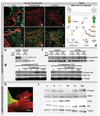
BMP signaling and FGF signaling at the inflow of the heart. Immunofluorescent images of the proepicardium and flanking inflow myocardium showing, cTnI (a), P-Smad (b), and P-Erk1/2 (c). a′ through c′ show and overlay the respective patterns with the nuclei. d schematically shows the interaction of BMP signaling via Smad (1) and FGF signaling via Erk1/2 (2). Erk1/2 phosphorylation of P-Smad results in abrogation of BMP signaling attributable to cytoplasmic P-Smad accumulation and degradation., H10 cells have elevated levels of P-Erk, which can be inhibited by the Mek1/2 inhibitor U0126 (e). Phosphorylation of Smad on BMP2 stimulation is increased when Mek1/2 is inhibited (f). Pretreatment with FGF2 inhibits the phosphorylation of Smad on BMP2 stimulation (g). This interaction was also shown in freshly isolated proepicardial cells; the dashed line indicates the position of the section. h, BMP2 stimulated Smad phosphorylation, which is inhibited by simultaneous addition of FGF2. When Mek1/2 was inhibited, Erk1/2 was not phosphorylated on FGF stimulation. The inhibiting effect of FGF2 on Smad phosphorylation was uncoupled by addition of U0126 in combination with BMP2 + FGF2 (i).
Figure 4
Figure 4
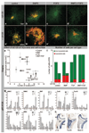
The effect of Mek1/2 inhibition on myocardium formation in vitro. Representative examples of control (a) BMP2- (b), FGF2-(c), and BMP2+FGF2-treated (d) proepicardial cultures in the absence (a through d) or presence of U0126 (a′ through d′) after 5 days of culture in which myocardium is visualized in red (cTnI) and nuclei in green. e, The graph shows the changes in the area of myocardium (y axis) and the number of nuclei (x axis) in each of the conditions (mean±SEM). Inhibition of Mek1/2 (gray arrows) decreases the number of cells without affecting the myocardial area in control and BMP2-treated cultures, whereas in FGF2-and BMP2 + FGF2-treated cultures the myocardial area is increased without affecting the total cell number. f, The bar graph shows the effect of the treatments on the number of myocardial and nonmyocardial cells. Parameters that are not significantly different are represented at the level of their geometric mean value. In control and BMP2-treated cultures, U0126 affects the number of nonmyocardial cells without affecting the number of myocardial cells, whereas U0126 increases the number of myocardial cells in FGF2 and BMP2+FGF2-treated cultures. g, Cultured proepicardia were analyzed by quantitative RT-PCR and compared to myocardial and epicardial cells. SV indicates sinus venosus; A, atrium; V, ventricle. h shows the expression of Tbx18, Nkx2.5, and cTnI in the proepicardium and flanking sinus venosus.
Figure 5
Figure 5
Twenty-four-hour treatment of developing embryos. In embryos treated with U0126+BMP2 for 24 hour before euthanasia, the proepicardium is a small sac-like structure (b) compared to controls (arrow heads) (a), and in the base of the rudimentary proepicardium, myocardial strands are present (red arrow) (b). In FGF2-treated embryos, an epicardium has already started to develop (black arrow) (c). In control and U0126+BMP2-treated embryos, proliferation is similar as assessed by 5-bromodeoxyuridine incorporation (red) (d and f). TUNEL assays showed no apoptotic cells in the proepicardium of control (e) or U0126+BMP2-treated embryos (g).
Figure 6
Figure 6
Forty-eight-hour treatment of developing embryos. In embryos treated with U0126+BMP2 for 48 hours before euthanasia, the epicardium is absent (b′), whereas in control embryos, almost the entire myocardium is covered with epicardium (a′). In FGF-treated embryos proepicardium formation takes place in a larger area of the venous pole of the heart, which coincides with smaller myocardial sleeves covering the sinus horns (c′ and f). Three-dimensional reconstructions of the heart of a control (d) and U0126+BMP2-treated (e) embryo shows that the epicardium is absent and the inflow myocardium is more extensive in U0126+BMP2-treated embryos. At the outflow pole the myocardial border is located closer to the aortic arch arteries (arrow head). The dashed lines in the 3D reconstructions indicate the position of the sections shown in a through c. (See also Online 3D PDF).
Figure 7
Figure 7
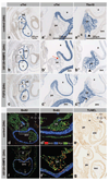
Model of separation of epicardial and myocardial cells from progenitors by BMP2 and FGF2. Balanced BMP2 + FGF2 signaling drives proliferation of progenitors. When the balance shifts in favor of FGF signaling via Erk1/2, the progenitors differentiate into epicardial cells. Shifting the balance in favor of BMP signaling via Smad results in myocardial differentiation. BMP signaling is inhibited by FGF signaling via Erk, which leads to cytoplasmic accumulation and degradation of P-Smad (a). A similar interaction of BMP and FGF signaling might still be operational in epicardium-derived cells in the formed heart (e) because cytoplasmic P-Smad (c) and P-Erk (d) are present in the subepicardial mesenchyme. The dashed line highlights the boundary between myocardium and epicardium based on cTnI staining (b and b′). b′ through d′ show an overlay of all nuclei. e shows the position of the enlargements (b through d) in the heart.

Comment in

References

    1. van den Berg G, Abu-Issa R, de Boer BA, Hutson MR, de Boer PA, Soufan AT, Ruijter JM, Kirby ML, van den Hoff MJ, Moorman AF. A caudal proliferating growth center contributes to both poles of the forming heart tube. Circ Res. 2009;104:179–188. - PMC - PubMed
    1. van den Hoff MJB, Kruithof BPT, Moorman AFM. Making more heart muscle. Bioessays. 2004;26:248–261. - PubMed
    1. Vliegen HW, van der LA, Cornelisse CJ, Eulderink F. Myocardial changes in pressure overload-induced left ventricular hypertrophy. A study on tissue composition, polyploidization and multinucleation. Eur Heart J. 1991;12:488–494. - PubMed
    1. Munoz-Chapuli R, Macias D, Gonzalez-Iriarte M, Carmona R, Atencia G, Perez-Pomares JM. The epicardium and epicardial-derived cells: multiple functions in cardiac development. Rev Esp Cardiol. 2002;55:1070–1082. - PubMed
    1. Cai CL, Martin JC, Sun Y, Cui L, Wang L, Ouyang K, Yang L, Bu L, Liang X, Zhang X, Stallcup WB, Denton CP, McCulloch A, Chen J, Evans SM. A myocardial lineage derives from Tbx18 epicardial cells. Nature. 2008;454:104–108. - PMC - PubMed

Publication types

MeSH terms