. 2009 Jul 24;325(5939):471-3.
doi: 10.1126/science.1173976.

Synchronous and stochastic patterns of gene activation in the Drosophila embryo

Affiliations

Synchronous and stochastic patterns of gene activation in the Drosophila embryo

Alistair N Boettiger et al. Science. .

Abstract

Drosophila embryogenesis is characterized by rapid transitions in gene activity, whereby crudely distributed gradients of regulatory proteins give way to precise on/off patterns of gene expression. To explore the underlying mechanisms, a partially automated, quantitative in situ hybridization method was used to visualize expression profiles of 14 developmental control genes in hundreds of embryos. These studies revealed two distinct patterns of gene activation: synchronous and stochastic. Synchronous genes display essentially uniform expression of nascent transcripts in all cells of an embryonic tissue, whereas stochastic genes display erratic patterns of de novo activation. RNA polymerase II is "pre-loaded" (stalled) in the promoter regions of synchronous genes, but not stochastic genes. Transcriptional synchrony might ensure the orderly deployment of the complex gene regulatory networks that control embryogenesis.


Figures

Fig. 1
Fig. 1
Assaying asynchrony of gene expression. (A to D) Nascent transcripts tagged with fluorescent probes (green) in whole-mount embryos allow for the activation state of each nucleus (purple) to be determined. All embryos are oriented anterior to the left, dorsal side to the top. The diagram above each image shows the gene prediction models (blue) and Pol II ChIP-chip binding data in dorsal-ectoderm (orange), neural-ectoderm (yellow), or mesodermal (red) tissue (37). htl and Mes4 are simultaneously expressed in the mesoderm [(A) and (B)]. Neu3 and ths are simultaneously expressed in the neural-ectoderm [(C) and (D)]. Genes that show a peak Pol II binding in the ChIP-chip data at the promoter in all tissues regulate transcriptional elongation via stalled polymerase [(A) and (C)]. Genes that lack promoter proximal Pol II binding in inactive tissue are nonstalled [(B) and (D)]. Gene names shown in black are stalled; purple are nonstalled.
Fig. 2
Fig. 2
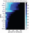
Asynchronous expression among paused and unpaused ectodermal. Heat map summarizing synchrony results for each gene. The color indicates the fraction of activated nuclei in the region of expression. The x axis shows the proportion of similarly staged embryos with a fractional activation of at least n [0,1]. The results are sorted from top to bottom by the sum along the row, putting the most asynchronous genes at the top and the most synchronous at the bottom.
Fig. 3
Fig. 3
Cell location and transcription factor gradient do not dramatically affect synchrony. (A and B) Embryos containing an anterior-posterior gradient of Dorsal activity due to toll[10b]-bcd-3′UTR mRNA being expressed in the anterior pole under control of the maternal hsp83 driver (24) were assayed for asynchronies in gene expression. The ectopic activation under control of a differently shaped dorsal gradient still recapitulates the WT observation that Fig. 2.
Fig. 4
Fig. 4
Robustness to perturbation. (A) The neurogenic ectoderm gene rho activates in a more stochastic fashion in Dorsal heterozygotes (dl/+) than in WT embryos [panel (C) and fig. S2]. (B) The neurogenic gene vnd shows no detectable difference in the synchrony of expression in the dl/+ background. (C) Summary of quantification of asynchronous expression for lateral ectoderm genes in WT (yw) and dl/+ embryos. Underlined gene names have confirmed shadow enhancers (29) and exhibit robust synchronous activation in the heterozygote background.

Similar articles

Cited by

References

    1. Raj A, Peskin CS, Tranchina D, Vargas DY, Tyagi S. PLoS Biol. 2006;4:e309. - PMC - PubMed
    1. Raj A, van Oudenaarden A. Cell. 2008;135:216. - PMC - PubMed
    1. Raser JM, O'Shea EK. Science. 2004;304:1811. published online 27 May 2004 (10.1126/science.1098641) - PMC - PubMed
    1. Raser JM, O'Shea EK. Science. 2005;309:2010. - PMC - PubMed
    1. Hendrix DA, Hong JW, Zeitlinger J, Rokhsar DS, Levine MS. Proc. Natl. Acad. Sci. U.S.A. 2008;105:7762. - PMC - PubMed

Publication types

Substances