. 2009 Sep 18;284(38):25842-53.
doi: 10.1074/jbc.M109.043208. Epub 2009 Jul 23.

Investigating the elusive mechanism of glycosaminoglycan biosynthesis

Affiliations

Investigating the elusive mechanism of glycosaminoglycan biosynthesis

Xylophone V Victor et al. J Biol Chem. .

Abstract

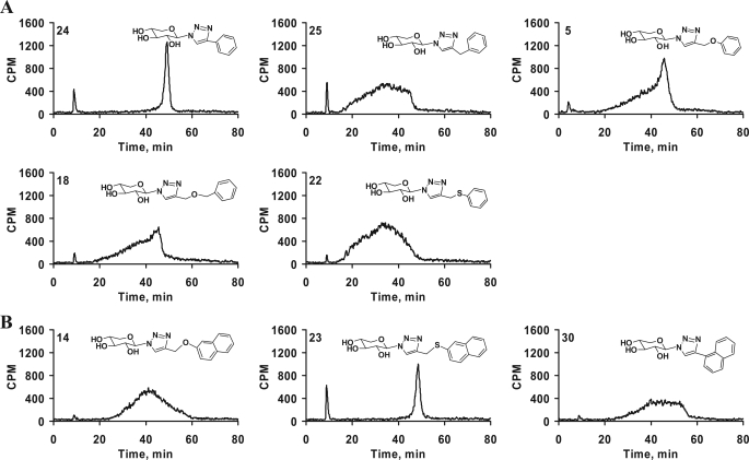
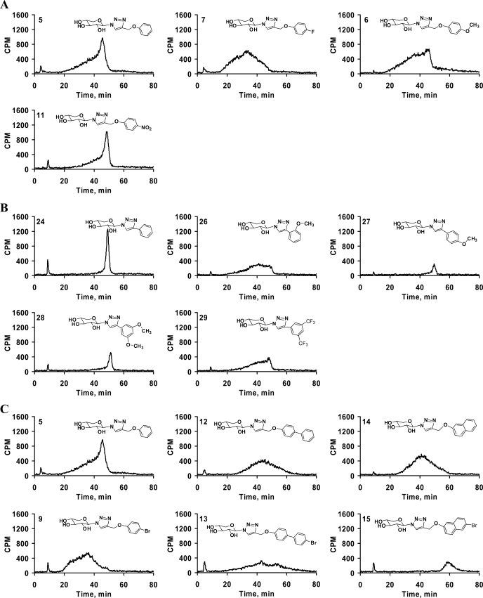
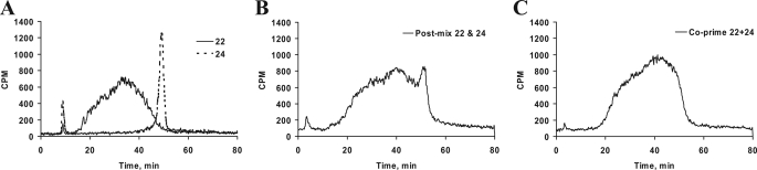
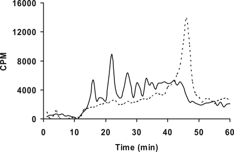
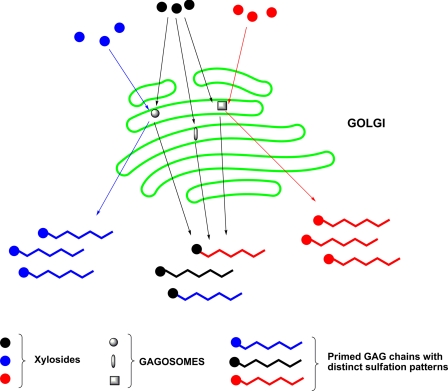
Glycosaminoglycan (GAG) biosynthesis requires numerous biosynthetic enzymes and activated sulfate and sugar donors. Although the sequence of biosynthetic events is resolved using reconstituted systems, little is known about the emergence of cell-specific GAG chains (heparan sulfate, chondroitin sulfate, and dermatan sulfate) with distinct sulfation patterns. We have utilized a library of click-xylosides that have various aglycones to decipher the mechanism of GAG biosynthesis in a cellular system. Earlier studies have shown that both the concentration of the primers and the structure of the aglycone moieties can affect the composition of the newly synthesized GAG chains. However, it is largely unknown whether structural features of aglycone affect the extent of sulfation, sulfation pattern, disaccharide composition, and chain length of GAG chains. In this study, we show that aglycones can switch not only the type of GAG chains, but also their fine structures. Our findings provide suggestive evidence for the presence of GAGOSOMES that have different combinations of enzymes and their isoforms regulating the synthesis of cell-specific combinatorial structures. We surmise that click-xylosides are differentially recognized by the GAGOSOMES to generate distinct GAG structures as observed in this study. These novel click-xylosides offer new avenues to profile the cell-specific GAG chains, elucidate the mechanism of GAG biosynthesis, and to decipher the biological actions of GAG chains in model organisms.


Figures

FIGURE 1.
FIGURE 1.
Priming activity of various xylosides. The novel xylosides were examined for their priming ability using xylosyl transferase-deficient CHO cells (pgsA-745). 100,000 cells were seeded per well of 24-well plates and treated with various xylosides at 0.1, 1, 10, and 100 μm and 1 mm concentrations for 24 h in the presence of 50 μCi of [35S]O42− or d-[6-3H]glucosamine. The GAG chains were then purified and quantified as described under “Experimental Procedures.” Xylosides with different aglycones primed different amounts of GAG chains at different concentrations suggesting the decisive role played by aglycones. The data indicate the average of three independent experiments.
FIGURE 2.
FIGURE 2.
Effect of substituents and their positions on priming activity. Cells were treated with various xylosides for 24 h in the presence of 50 μCi of [35S]O42− or d-[6-3H]glucosamine as described under “Experimental Procedures.” Primed GAG chains were then purified from the supernatant using anion-exchange column chromatography and quantified using liquid scintillation. A, effect of various halogen substituents on the priming activity was examined. Fluoro-, chloro-, bromo-, and iodo-substituted xylosides (7, 8, 9, and 10, respectively) were compared against the unsubstituted xyloside 5 at 1 mm concentration. B, effect of methoxy-substituent and its position on the phenyl ring was examined for priming activity. Unsubstituted xyloside 24 was compared with ortho-substituted xyloside 26 and para-substituted xyloside 27 at 100 μm (unshaded) and 1 mm (shaded) concentrations. The data indicate the average of three independent experiments.
FIGURE 3.
FIGURE 3.
Correlation between extent of sulfation and migration time on HPLC anion-exchange chromatography. Cells containing d-[6-3H]glucosamine were treated with 100 μm xyloside 5 in the presence of chlorate at various concentrations. GAG chains were then isolated and quantified as described under “Experimental Procedures.” The chlorate treatment did not affect the amount of GAG chains produced. 1,000,000 cpm were applied to HPLC DEAE anion-exchange chromatography and eluted with a linear NaCl gradient as described under “Experimental Procedures.” GAG species from chlorate-treated cells were eluted earlier than those from control cells. The elution profiles of GAG chains from control cells (gray tracer), cells treated with 5 mm chlorate (dark tracer), and cells treated with 25 mm chlorate (broken tracer) are shown. The elution profiles are representative of at least two independent experiments.
FIGURE 4.
FIGURE 4.
Effect of various hydrophobic moieties on the DEAE elution profile of GAG chains. GAG chains primed by various xylosides (1, 3, 5, 12, 14, and 17) eluted from small anion-exchange column with 1 m NaCl, were diluted 5-fold and analyzed by anion-exchange HPLC column as described under “Experimental Procedures.” The variations in the elution profiles and migration times indicate differences in both sulfation pattern and extent of sulfation of the primed GAG chains. The elution profiles are representative of at least two independent experiments.
FIGURE 5.
FIGURE 5.
Spacers between the triazole and aromatic ring in the aglycone moiety alter the sulfation pattern and extent of sulfation. Purified GAG chains were diluted 5-fold and analyzed by anion-exchange HPLC column as described under “Experimental Procedures.” A gradient of 0.2 to 1.0 m NaCl for 80 min was used for elution of the GAG chains. The differential elution profile and migration time indicates variations in sulfation pattern and extent of sulfation of GAG chains primed by phenyl group containing xylosides 5, 18, 22, 24, and 25 (A) and naphthyl group containing xylosides 14, 23, and 30 having various spacers were examined (B). The elution profiles are representative of at least two independent experiments.
FIGURE 6.
FIGURE 6.
Effect of substituents and their positions on GAG fine structures. Purified GAG chains were diluted 5-fold and analyzed by HPLC DEAE anion-exchange chromatography as described under “Experimental Procedures.” The differential elution profiles and migration times indicate variations in sulfation pattern and extent of sulfation. A, effect of various substituents at the para position of the phenyl ring was examined. The elution suggests that different substituents on xylosides, 5, 6, 7, and 11 affect the elution profile of the GAG chains suggesting changes in the fine structures. B, influence of position of the substituent on the fine structures was examined. Unsubstituted xyloside 24 primed GAG chains that elute as a narrow peak, whereas ortho methoxy-substituted xyloside 26 produced GAG chains that elute as a broad peak. On the other hand, para-substituted xyloside 27 and dimethoxy-substituted xyloside 28 produce GAG chains that elute as narrow peaks. C, bromo-substitution on phenyl, biphenyl, and naphthyl moieties was found to affect the extent of sulfation and sulfation pattern. Unsubstituted xylosides 5, 12, and 14 were compared with their corresponding bromo-substituted xylosides 9, 13, and 15 for the change in the extent of sulfation and sulfation pattern. The elution profiles are representative of at least two independent experiments.
FIGURE 7.
FIGURE 7.
Co-priming experiment with xylosides that prime distinct GAG chains. A, xylosides 22 and 24 were primed at 100 μm concentration in pgsA-745 cells for 24 h individually, and GAG chains were purified. Individually primed GAG chains were then analyzed on HPLC DEAE anion-exchange chromatography under similar conditions in separate runs. The solid tracer indicates GAG chains primed by xyloside 22, and the broken tracer indicates the narrow elution profiles of the GAG chains primed by xyloside 24. B, individually primed GAG chains from xylosides 22 and 24 were mixed together and analyzed under similar conditions as described in A. C, 100 μm each of xylosides 22 and 24 were primed together for 24 h in cell culture experiments. The resulting GAG chains were purified and analyzed by DEAE anion-exchange chromatography as described earlier. The elution profile suggests further loss of resolution of two distinct GAG chains when xylosides 22 and 24 are co-primed. The elution profiles are representative of at least two independent experiments.
FIGURE 8.
FIGURE 8.
Chondroitinase B treatment of GAG chains primed by xyloside 20. Xyloside 20 was primed at 100 μm concentration in pgsA-745 cells for 24 h. The GAG chains were then purified as described under “Experimental Procedures.” The purified GAG chains were digested with chondroitinase B, analyzed by HPLC DEAE anion-exchange chromatography, and compared with the undigested GAG chains. The elution profiles of the undigested (broken tracer) and digested (solid tracer) GAG chains are shown.
FIGURE 9.
FIGURE 9.
Regulation of GAG biosynthesis by GAGOSOME model. Each GAGOSOME can have different combination of enzymes that generate cell-specific combinatorial GAG structures with differential sulfation pattern required for binding to diverse proteins. While some xylosides (blue and red) are selectively primed by a specific GAGOSOME to generate distinct fine structures, other xylosides (black) are promiscuously primed by more than one GAGOSOME resulting in heterogeneous GAG chains.

Similar articles

Cited by

References

    1. Sasisekharan R., Shriver Z., Venkataraman G., Narayanasami U. (2002) Nat. Rev. Cancer 2, 521–528 - PubMed
    1. Lander A. D. (1993) Curr. Opin. Neurobiol. 3, 716–723 - PubMed
    1. Capila I., Linhardt R. J. (2002) Angew. Chem. Int. Ed. Engl. 41, 391–412 - PubMed
    1. Powell A. K., Yates E. A., Fernig D. G., Turnbull J. E. (2004) Glycobiology 14, 17R–30R - PubMed
    1. Salmivirta M., Lidholt K., Lindahl U. (1996) FASEB J. 10, 1270–1279 - PubMed

Publication types

LinkOut - more resources