. 2009 Sep 18;284(38):25593-601.
doi: 10.1074/jbc.M109.025452. Epub 2009 Jul 23.

Angiopoietin-like 4 (ANGPTL4, fasting-induced adipose factor) is a direct glucocorticoid receptor target and participates in glucocorticoid-regulated triglyceride metabolism

Affiliations

Angiopoietin-like 4 (ANGPTL4, fasting-induced adipose factor) is a direct glucocorticoid receptor target and participates in glucocorticoid-regulated triglyceride metabolism

Suneil K Koliwad et al. J Biol Chem. .

Erratum in

  • J Biol Chem. 2012 Feb 3;287(6):4394

Abstract

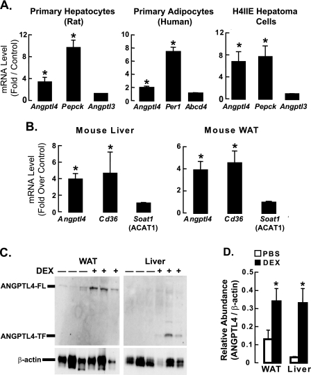
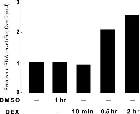
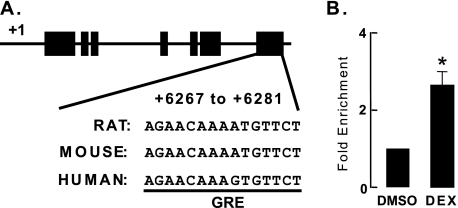
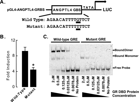
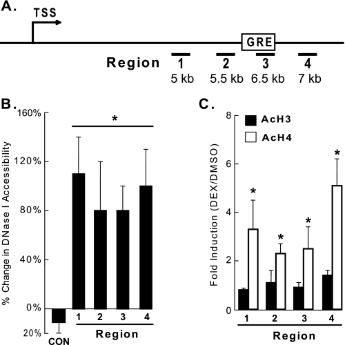
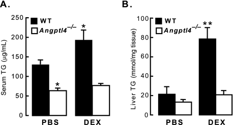
Glucocorticoids are important regulators of lipid homeostasis, and chronically elevated glucocorticoid levels induce hypertriglyceridemia, hepatic steatosis, and visceral obesity. The occupied glucocorticoid receptor (GR) is a transcription factor. However, those genes regulating lipid metabolism under GR control are not fully known. Angiopoietin-like 4 (ANGPTL4, fasting-induced adipose factor), a protein inhibitor of lipoprotein lipase, is synthesized and secreted during fasting, when circulating glucocorticoid levels are physiologically increased. We therefore tested whether the ANGPTL4 gene (Angptl4) is transcriptionally controlled by GR. We show that treatment with the synthetic glucocorticoid dexamethasone increased Angptl4 mRNA levels in primary hepatocytes and adipocytes (2-3-fold) and in the livers and white adipose tissue of mice (approximately 4-fold). We tested the mechanism of this increase in H4IIE hepatoma cells and found that dexamethasone treatment increased the transcriptional rate of Angptl4. Using bioinformatics and chromatin immunoprecipitation, we identified a GR binding site within the rat Angptl4 sequence. A reporter plasmid containing this site was markedly activated by dexamethasone, indicative of a functional glucocorticoid response element. Dexamethasone treatment also increased histone H4 acetylation and DNase I accessibility in genomic regions near this site, further supporting that it is a glucocorticoid response element. Glucocorticoids promote the flux of triglycerides from white adipose tissue to liver. We found that mice lacking ANGPTL4 (Angptl4(-/-)) had reductions in dexamethasone-induced hypertriglyceridemia and hepatic steatosis, suggesting that ANGPTL4 is required for this flux. Overall, we establish that ANGPTL4 is a direct GR target that participates in glucocorticoid-regulated triglyceride metabolism.


Figures

FIGURE 1.
FIGURE 1.
ANGPTL4 gene and protein expression are regulated by glucocorticoids in vivo and in vitro. A, rat primary hepatocytes, human primary adipocytes, and rat H4IIE hepatoma cells (n = 3–5) were treated with either DMSO or DEX (0.5 μm) for 5 h, after which mRNA levels of Angptl4 and positive and negative control genes were measured by qPCR. Data show fold-induction of gene expression (DEX/DMSO) from at least three separate experiments (*, p < 0.05). B, mice (n = 6) were treated with either PBS or DEX (40 mg/kg) for 4 days then fasted for 4 h, after which their livers and epididymal fat (WAT) were harvested to perform qPCR as in A (*, p < 0.05). C, liver and WAT samples from control and DEX-treated mice in B were also collected for analysis of protein expression by Western blot (6.25 μg of tissue per sample run for WAT and 100 μg for liver). For each blot, the first 3 lanes are samples from individual mice treated with PBS (−), and the last 3 lanes are from DEX-treated mice (+). Full-length ANGPTL4 (ANGPTL4-FL) was the main band in WAT, whereas the truncated form (ANGPTL4-TF) predominated in the liver. β-Actin served as the internal loading control. D, quantification of the intensity of ANGPTL4 bands (see “Experimental Procedures”) from Western blots as in C (n = 3). Data represent relative optical density (ANGPTL4/β-actin; *, p < 0.05). The error bars represent the S. E. for the fold induction and the relative abundance.
FIGURE 2.
FIGURE 2.
DEX treatment increases the rate of Angptl4 gene transcription in H4IIE cells. H4IIE cells were untreated, treated with DMSO for 120 min, or with DEX (0.5 μm) for 10, 30, or 120 min as shown. Nuclei from these cells were used for in vitro transcription with biotin UTP. Newly synthesized RNA was isolated on streptavidin beads. cDNA was then synthesized and qPCR performed to monitor for changes in transcription rates using primers specific to Angptl4 and β-actin (control). Shown is the fold-induction in transcription of DEX-treated samples (DEX/untreated) from one of two independent experiments showing similar results.
FIGURE 3.
FIGURE 3.
Identification of an Angptl4 GRE. A, schematic diagram of rat Angptl4, including the location of the predicted GR binding site. Black boxes represent exons, and lines connecting them represent introns. The predicted GR binding site sequences (AGAACATTTTGTTCT) and their conserved counterparts in human and mouse genome are shown. B, ChIP experiments confirming the recruitment of GR to its predicted binding site. H4IIE cells were treated with either DMSO or DEX (0.5 μm) for 30 min, after which they were harvested for ChIP as described (“Experimental Procedures”). Shown is the fold-enrichment of GR binding by DEX treatment (DEX/DMSO) from three independent experiments (*, p < 0.05). The error bars represent the S. E. for the fold enrichment.
FIGURE 4.
FIGURE 4.
The GR binding region of Angptl4 confers glucocorticoid responsiveness. A, a sequence containing either the wild-type or a mutated Angptl4 GR binding site was inserted into the pGL4-TATA reporter to create pGL4-ANGPTL4-GRBS (mutation from Gly to Thr underlined). B, glucocorticoid responsiveness of GR-binding sites in rat Angptl4. Reporter plasmids and wild-type or mutant pGL4-ANGPTL4-GRBS (75 ng) were cotransfected with pcDNA3-hGR (150 ng) and pRL (100 ng) into H4IIE cells in a 24-well plate (n = 4 per group). pRL plasmid provided Renilla luciferase expression to document transfection efficiency. Transfected cells were left overnight, then washed with PBS and treated with 0.5 μm DEX for an additional 16–20 h. Cells were then lysed and assayed for firefly and Renilla luciferase activities. Shown is the fold-induction of luciferase activity (DEX-treated/ethanol-treated) in cells from at least three experiments (*, p < 0.05). The error bars represent the S. E. for the fold induction. C, EMSA on mixtures of purified GR DBD and Cy5 end-labeled oligonucleotides containing either wild-type or mutant Angptl4 GRE, confirming direct binding of GR to the GRE. Reactions lasted 30 min, and the mixtures were then run on 8% native gels and scanned by a phosphorimager. The concentration of GR DBD protein mixed with wild-type or mutant GREs ranged from 0 (no protein) to 2 μm, as shown. Data are representative of two independent experiments.
FIGURE 5.
FIGURE 5.
DEX treatment increases DNase I accessibility and histone H4 acetylation in rat Angptl4. A, schematic diagram of rat Angptl4, with TSS indicating the transcriptional start site, amplified genomic regions are underlined and numbered (–4), and the position of the GRE shown. B, H4IIE cells were treated with DMSO or DEX (0.5 μm) for 30 min, and DNase I accessibility was analyzed as described (“Experimental Procedures”), showing increased cleavage of DNA in DEX-treated cells. C, H4IIE cells (n = 4 per group) were treated with DMSO or DEX (0.5 μm) for 30 min, and the levels of acetylated histones H3 and H4 were measured in regions 1–4 by ChIP. The fold-enrichment (DEX/DMSO) was used to show increased enrichment of AcH4 but not AcH3 in DEX-treated cells (*, p < 0.05). Data in B and C were normalized to a control genomic region in rat Rpl19, and each experiment was done at least 3 times. The error bars represent the S. E. for the percent change and the fold induction.
FIGURE 6.
FIGURE 6.
The DEX-stimulated increase in serum and liver TG is impaired in Angptl4−/− mice. A, wild-type and Angptl4−/− mice were treated daily with PBS (control; n = 4–5 per group) or DEX (n = 5 per group) for 4 days as described (“Experimental Procedures”), after which serum TG levels were measured. Shown is the fold-increase in serum TG of DEX-treated mice (*, p < 0.05 versus WT PBS) for both wild-type and Angptl4−/− mice. B, liver TG content measured by TLC from the same mice as in A (**, p < 0.05 versus WT PBS). The error bars represent the S. E. for the TG concentration.

Similar articles

Cited by

References

    1. Macfarlane D. P., Forbes S., Walker B. R. (2008) J. Endocrinol. 197, 189–204 - PubMed
    1. Seckl J. R., Morton N. M., Chapman K. E., Walker B. R. (2004) Recent Prog. Horm. Res. 59, 359–393 - PubMed
    1. Hato T., Tabata M., Oike Y. (2008) Trends Cardiovasc. Med. 18, 6–14 - PubMed
    1. Kersten S. (2005) Biochem. Soc. Trans. 33, 1059–1062 - PubMed
    1. Li C. (2006) Curr. Opin. Lipidol. 17, 152–156 - PubMed

Publication types

MeSH terms