

Mutations in mitochondrial DNA polymerase-gamma promote breast tumorigenesis
- PMID: 19629138
- PMCID: PMC2782392
- DOI: 10.1038/jhg.2009.71
Mutations in mitochondrial DNA polymerase-gamma promote breast tumorigenesis
Abstract
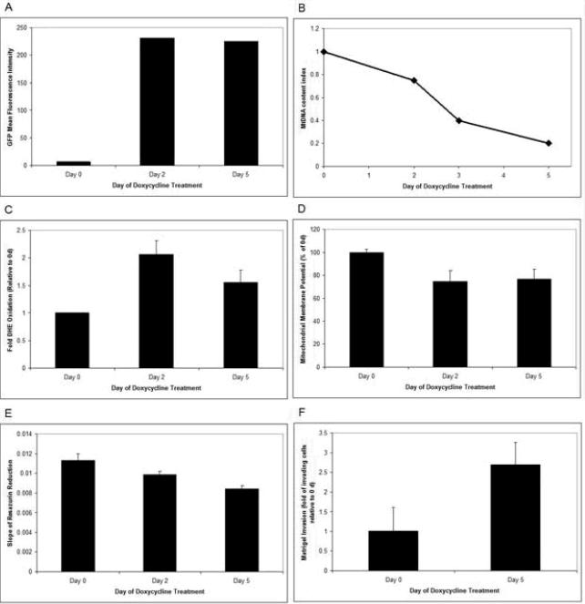
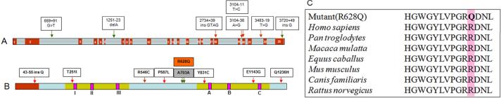
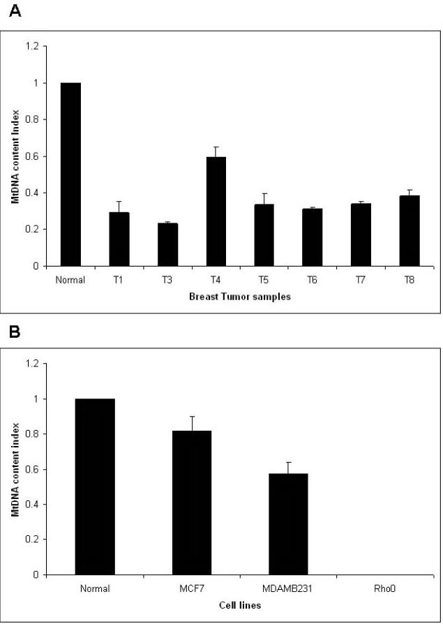
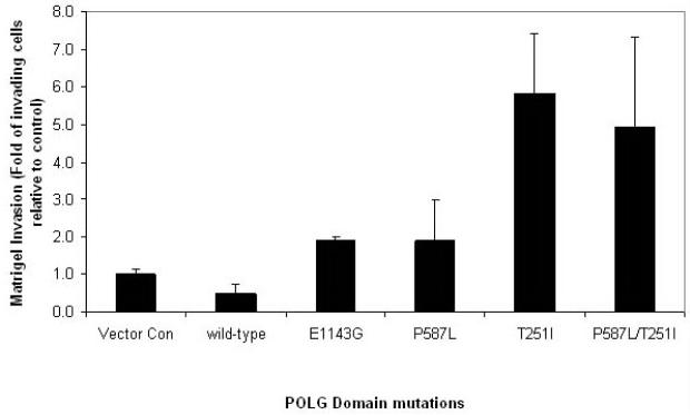
Decreased mitochondrial oxidative phosphorylation (OXPHOS) is one of the hallmarks of cancer. To date, the identity of nuclear gene(s) responsible for decreased OXPHOS in tumors remains unknown. It is also unclear whether mutations in nuclear gene(s) responsible for decreased OXPHOS affect tumorigenesis. Polymerase-gamma (POLG) is the only DNA polymerase known to function in human mitochondria. Mutations in POLG are known to cause mitochondrial DNA (mtDNA) depletion and decreased OXPHOS, resulting in mtDNA depletion syndrome in humans. We therefore sequenced all coding exons (2-23) and flanking intron/splice junctions of POLG in breast tumors. We found that the POLG gene was mutated in 63% of breast tumors. We identified a total of 17 mutations across the POLG gene. Mutations were found in all three domains of the POLG protein, including T251I (the exonuclease domain), P587L (the linker region) and E1143G (the polymerase domain). We identified two novel mutations that include one silent (A703A) and one missense (R628Q) mutation in the evolutionarily conserved POLG linker region. In addition, we identified three novel mutations in the intronic region. Our study also revealed that mtDNA was depleted in breast tumors. Consistently, mutant POLG, when expressed in breast cancer cells, induced a depletion of mtDNA, decreased mitochondrial activity, decreased mitochondrial membrane potential, increased levels of reactive oxygen species and increased Matrigel invasion. Together, our study provides the first comprehensive analysis of the POLG gene mutation in human cancer and suggests a function for POLG (1) in decreased OXPHOS in cancers and (2) in promoting tumorigenicity.
Figures
References
-
- Warburg O. Metabolism of Tumors. Arnold Constable; London, UK: 1930.
-
- Warburg O. On respiratory impairment in cancer cells. Science. 1956;124:269–270. - PubMed
-
- Pedersen PL. Bioenergetics of cancer cells. J Bioenerg Biomembr. 1997;29:301–302. - PubMed
-
- Pedersen PL. Warburg, me and Hexokinase 2: Multiple discoveries of key molecular events underlying one of cancers' most common phenotypes, the “Warburg Effect”. J Bioenerg Biomembr. 2007;39:349–355. - PubMed
-
- Singh KK. Mitochondrial DNA mutations in Aging, Disease, and Cancer. Springer; New York, USA: 1998.
Publication types
MeSH terms
Substances
Grants and funding
LinkOut - more resources
Full Text Sources
Medical