. 2009 Jul;5(7):e1000573.
doi: 10.1371/journal.pgen.1000573. Epub 2009 Jul 24.

Circadian clock genes contribute to the regulation of hair follicle cycling

Affiliations

Circadian clock genes contribute to the regulation of hair follicle cycling

Kevin K Lin et al. PLoS Genet. 2009 Jul.

Abstract

Hair follicles undergo recurrent cycling of controlled growth (anagen), regression (catagen), and relative quiescence (telogen) with a defined periodicity. Taking a genomics approach to study gene expression during synchronized mouse hair follicle cycling, we discovered that, in addition to circadian fluctuation, CLOCK-regulated genes are also modulated in phase with the hair growth cycle. During telogen and early anagen, circadian clock genes are prominently expressed in the secondary hair germ, which contains precursor cells for the growing follicle. Analysis of Clock and Bmal1 mutant mice reveals a delay in anagen progression, and the secondary hair germ cells show decreased levels of phosphorylated Rb and lack mitotic cells, suggesting that circadian clock genes regulate anagen progression via their effect on the cell cycle. Consistent with a block at the G1 phase of the cell cycle, we show a significant upregulation of p21 in Bmal1 mutant skin. While circadian clock mechanisms have been implicated in a variety of diurnal biological processes, our findings indicate that circadian clock genes may be utilized to modulate the progression of non-diurnal cyclic processes.


Conflict of interest statement

The authors have declared that no competing interests exist.

Figures
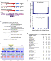
Figure 1
Figure 1. Identification of hair cycle–regulated genes using probabilistic models.
(A) Representative time points selected for gene expression profiling of the natural and depilation-induced hair growth cycles. Time points for natural cycles represent postnatal days, and time points for depilation-induced cycle indicate the number of days following depilation. Since the three cycles have different duration, the schematic timeline is not in actual time scale. Bottom panel consists of representative histology of dorsal skin at key phases of synchronized hair follicle cycling. (B) Overview of data processing and statistical analyses. Note that samples for the first synchronized hair growth cycle and asynchronous time points were profiled in an earlier study using an older generation array (Affymetrix Murine Genome U74Av2). (C) Schematic of the probabilistic model for detection of periodic gene expression changes during hair follicle cycling. See Materials and Methods for a detailed description of the schematic. Asynch – asynchronous cycle. (D) Histogram of the number of probe sets within each range of the indicated posterior probabilities of being periodically expressed. Top panel is the histogram of probabilities for all present genes during the hair growth cycle. The bottom panel is the histogram of probabilities for the literature-based hair cycle-dependent genes. (E) List of significantly enriched GO Biological Process categories within the sets of genes specifically upregulated at the indicated phases of the hair growth cycle. Due to the redundancy of categories, not all are listed. The number of genes upregulated at each phase of the cycle is in parentheses. Statistical enrichment of each category is shown as P-values calculated using a modified Fisher Exact test (DAVID functional annotation analysis).
Figure 2
Figure 2. CLOCK–regulated genes are periodically expressed during hair follicle cycling.
(A) Temporal clusters (labeled 1–6 and color coded) of hair cycle-regulated transcriptional regulators. Transcription factors that play key roles in hair follicle morphogenesis and/or cycling are labeled with gene symbols in black. CLOCK-regulated genes are labeled in the blue box. Expression levels are from profiling data of the second hair growth cycle and are indicated by the colorimetric ratio-scale. (B) Time-course profiles of CLOCK-controlled genes during hair follicle morphogenesis, the first two natural and depilation-induced hair growth cycles. For each gene, the expression levels were normalized relative to the lowest expressed time point of the second cycle; the first cycle (P1 to P23) was normalized separately because different array was used for profiling. Differences in magnitude of change between the first and second cycles are primarily due to differential probe set efficiencies. Note the broken y-axis. E – embryonic days; P – postnatal days; D - depilation days. (C) Q-PCR of Dbp using independent samples from the first two synchronized hair growth cycles. Standard deviations were determined by using three replicates normalized to Gapdh and fold calculated relative to the lowest expression sample. For (B–D), time points are mapped based on histology to the corresponding phases of the cycle: hair follicle morphogenesis (M), anagen (A), catagen (C), and telogen (T).
Figure 3
Figure 3. Enhanced circadian expression of CLOCK–regulated genes during telogen.
Q-PCR of Bmal1 (A), Clock (B), Dbp (C), Per2 (D), and Nr1d1 (E) in telogen compared to late anagen dorsal skin over the course of 48 hours (open and filled bars along the x-axis denote 12 hours light and dark phases, respectively). Expression is normalized to Gapdh and fold calculated relative to the lowest expression time point for both telogen and late anagen. Each error bar represents the S.E.M. for independent measurements from four mice. Asterisks denote significantly higher (P<0.01) expression of Dbp and Nr1d1 at ZT10 in telogen.
Figure 4
Figure 4. Bmal1 and its target gene Dbp are co-expressed during hair follicle cycling.
In situ hybridization staining of telogen, early anagen, late anagen, and catagen dorsal skin at ZT10 with Bmal1 (A–D) and Dbp (E–H) probes. (I) Dbp expression at ZT2 in telogen skin. Dashed lines indicate border between epidermis and dermis. Note that the black pigment of the hair shaft in late anagen hair follicles is not hybridization signal. Bu – bulge, CH – club hair, DP – dermal papilla, HB – hair bulb, HS – hair shaft, IRS – inner root sheath, Mx – matrix, ORS – outer root sheath, SHG – secondary hair germ, SG – sebaceous gland. (J) Dbp expression from laser capture microdissected hair follicles, dermis, and epidermis for telogen and late anagen dorsal skin at ZT10 and ZT18/ZT2. Standard deviations were determined by using three replicates normalized to Gapdh. Ct values indicate detectable expression of clock genes in every sample, and fold was calculated relative to the lowest expression sample.
Figure 5
Figure 5. Significant differential expression of circadian transcriptional circuit and key cell cycle regulators in telogen Bmal1 and Clock mutant dorsal skin.
(A) Overview of microarray analysis of P22 Bmal1 −/− and Bmal1 +/− dorsal skin. FDR – false discovery rate. (B) The statistical differential expression of clock genes is shown with the circadian transcriptional circuit, which is represented as a matrix based on the results of Ueda et al. . In the matrix, each column represents a gene encoding a transcription factor (grouped into three classes based on their binding sites: E-box/E′-box, D-box, and RRE), and each row represents a gene that is regulated by these transcription factors. Orange cells denote positive regulation and blue cells denote negative regulation. (C) Q-PCR of CLOCK-regulated genes and key cell cycle regulators in P22 Bmal1 −/− and Bmal1 +/− dorsal skin. (D) Q-PCR of CLOCK-regulated genes in P23 Clock wild-type and Clock mutant dorsal skin. For (C) and (D), expression is normalized to Gapdh and asterisks denote statistically significant (P<0.01) difference in expression between the littermates of the two genotypes. Error bars represent the S.E.M. for independent measurements from five Bmal1 −/− and four Bmal1 +/− mice, and three Clock wild-type and two Clock mutant mice.
Figure 6
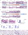
Figure 6. Bmal1 and Clock regulate anagen progression in hair follicle cycling.
(A) Representative histological sections of dorsal skin from Bmal1 −/− mice and their gender-matched Bmal1 +/− littermates at the indicated postnatal age (P). (B) Representative histological sections of dorsal skin from Clock mutant and their gender-matched wild-type (WT) littermates at the indicated postnatal age (P). For (A) and (B), time points are mapped (indicated by dotted line) based on histology to the corresponding phases of the hair growth cycle: anagen (A), catagen (C), and telogen (T). (C) Delayed anagen progression in Bmal1 −/− mice compared to normal progression in Bmal1 +/− littermates. At postnatal day 24, hair follicles are in anagen IIIb for the shown Bmal1 +/− dorsal skin section; matrix cells (black arrow) form the enlarged hair bulb and the dermal papilla (red arrow) is larger than a third of bulb diameter. Note that the bulb is located in the middle of the subcutaneous adipose layer. Hair follicles are in anagen I for the shown Bmal1 −/− dorsal skin section; thickening of keratinocyte strand (blue arrow) between the dermal papilla (red arrow) and the club hair. Note that the bulb is located in the dermis. Brackets indicate the different layers of the skin: E – epidermis, D – dermis, SC – subcutaneous adipose layer. (D) Quantitative hair cycle histomorphometric analysis. Percentage of hair follicles at the indicated hair cycle stage is based on staging fifty unique hair follicles for each genotype from three Bmal1 +/− and two Bmal1 −/− littermates at P24. Tel – telogen. Ana – anagen (Roman numerals indicate specific stages within anagen). (E) Q-PCR of Ccnd1, Ccnb1, and Myc in P24 Bmal1 −/− and Bmal1 +/− dorsal skin. Expression is normalized to Gapdh and error bars represent the S.E.M. for independent measurements from six Bmal1 +/− and two Bmal1 −/− littermates. Asterisks denote statistically significant (P<0.01) difference in expression between the two genotypes. Immunostaining of dorsal skin from P24 Bmal1 +/− (left panel), Bmal1 −/− (central panel) littermates using anti-phospho-histone H3 (F) and anti-phospho-Rb (Ser807/811) (G). The right panels of A and B are wild-type mice at P23 with hair follicles at equivalent stage of the hair growth cycle (anagen I) to the P24 Bmal1 −/− mice. The insets are higher magnification of the lower regions of hair follicles. Black arrowheads; cells stained positive in the epidermis. Black dashed line; border between epidermis and dermis. Red dashed line; hair follicle bulb. Red arrowhead; cells stained positive within the hair follicle.

Similar articles

Cited by

References

    1. Stenn KS, Paus R. Controls of hair follicle cycling. Physiol Rev. 2001;81:449–494. - PubMed
    1. Paus R, Cotsarelis G. The biology of hair follicles. N Engl J Med. 1999;341:491–497. - PubMed
    1. Muller-Rover S, Handjiski B, van der Veen C, Eichmuller S, Foitzik K, et al. A comprehensive guide for the accurate classification of murine hair follicles in distinct hair cycle stages. J Invest Dermatol. 2001;117:3–15. - PubMed
    1. Millar SE. Molecular mechanisms regulating hair follicle development. J Invest Dermatol. 2002;118:216–225. - PubMed
    1. Fuchs E, Merrill BJ, Jamora C, DasGupta R. At the roots of a never-ending cycle. Dev Cell. 2001;1:13–25. - PubMed

Publication types