

Muscle pain: mechanisms and clinical significance
- PMID: 19629211
- PMCID: PMC2696782
- DOI: 10.3238/artzebl.2008.0214
Muscle pain: mechanisms and clinical significance
Abstract
Introduction: Muscle pain is common, but the understanding of its causes is still patchy. This article addresses the mechanisms of some important types of muscle pain.
Methods: Selective literature review, predominantly of data derived from neuroanatomical and electrophysiological experiments on anesthetized rats.
Results: Muscle pain is evoked by specialized nerve endings (nociceptors). Important stimuli for muscle pain are adenosintriphosphate (ATP) and a low tissue pH. Excitation of muscle nociceptors leads to hyperexcitability of spinal sensory neurones (central sensitization). Low frequency activity in muscle nociceptors is sufficient to induce central sensitization.
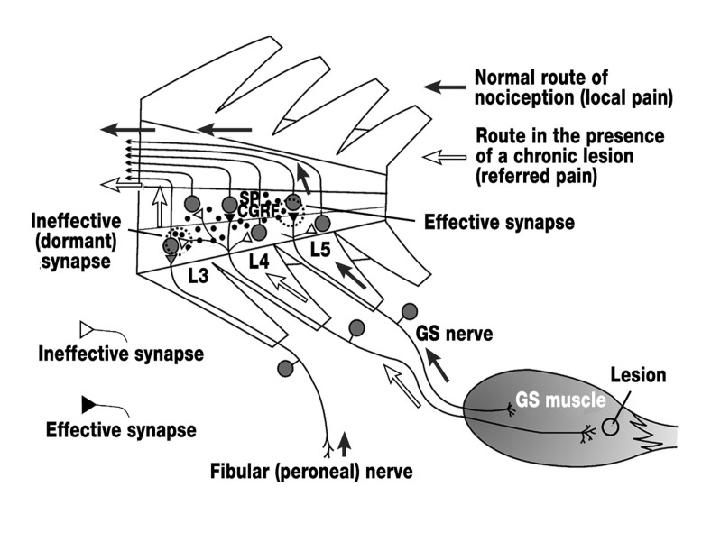
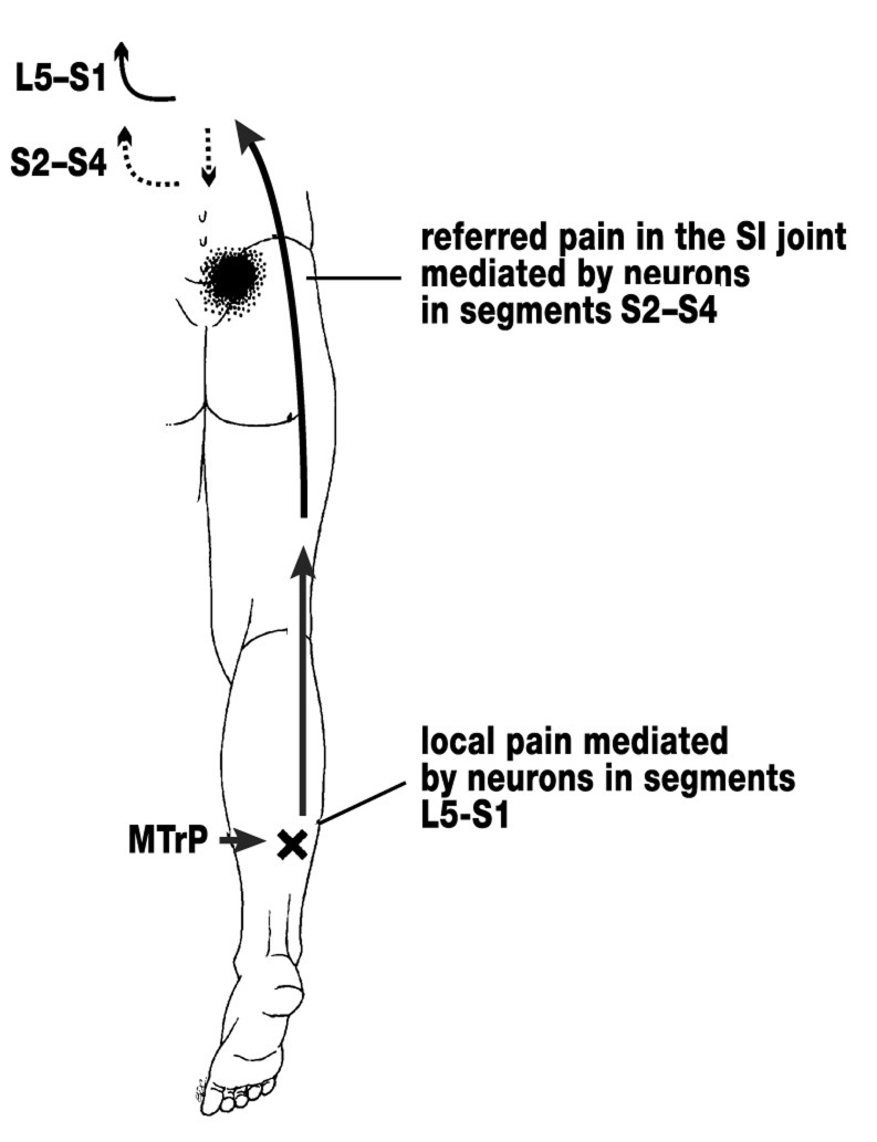
Discussion: Central sensitization leads to increased excitation in the spinal cord and to referral of muscle pain. The motoneurones of a painful muscle are centrally inhibited. Muscular spasm is mostly secondary to a painful lesion in another muscle or joint. The pain of fibromyalgia is assumed to relate to a dysfunction of central nociceptive processing. Psychosocial factors also contribute to pain.
Keywords: fibromyalgia; muscle pain; muscle spasm; myofascial trigger point; nociceptor; sensitization.
Figures
Comment in
-
The situation is more complex in patients with chronic pain.Dtsch Arztebl Int. 2008 Jul;105(28-29):510; author reply 510. doi: 10.3238/arztebl.2008.0510a. Epub 2008 Jul 14. Dtsch Arztebl Int. 2008. PMID: 19626205 Free PMC article. No abstract available.
-
Myofascial trigger points can be treated.Dtsch Arztebl Int. 2008 Jul;105(28-29):510; author reply 510. doi: 10.3238/arztebl.2008.0510b. Epub 2008 Jul 14. Dtsch Arztebl Int. 2008. PMID: 19626206 Free PMC article. No abstract available.
Similar articles
-
[Pathophysiology of low back pain and the transition to the chronic state - experimental data and new concepts].Schmerz. 2001 Dec;15(6):413-7. doi: 10.1007/s004820100002. Schmerz. 2001. PMID: 11793144 German.
-
Pathophysiological mechanisms of fibromyalgia.Clin J Pain. 1991;7 Suppl 1:S8-15. Clin J Pain. 1991. PMID: 1810527 Review.
-
[Neurobiological basis of muscle pain].Schmerz. 1999 Feb 18;13(1):3-17. doi: 10.1007/s004829900010. Schmerz. 1999. PMID: 12799945 German.
-
Algesic agents exciting muscle nociceptors.Exp Brain Res. 2009 Jun;196(1):89-100. doi: 10.1007/s00221-008-1674-4. Epub 2009 Jan 13. Exp Brain Res. 2009. PMID: 19139871
-
Central sensitization in fibromyalgia and other musculoskeletal disorders.Curr Pain Headache Rep. 2003 Oct;7(5):355-61. doi: 10.1007/s11916-003-0034-0. Curr Pain Headache Rep. 2003. PMID: 12946288 Review.
Cited by
-
Altered brain responses to noxious dentoalveolar stimuli in high-impact temporomandibular disorder pain patients.PLoS One. 2022 Oct 14;17(10):e0266349. doi: 10.1371/journal.pone.0266349. eCollection 2022. PLoS One. 2022. PMID: 36240243 Free PMC article.
-
Trigeminal neuralgia and chiropractic care: a case report.J Can Chiropr Assoc. 2010 Sep;54(3):177-86. J Can Chiropr Assoc. 2010. PMID: 20808617 Free PMC article.
-
Involvement of the Transient Receptor Channels in Preclinical Models of Musculoskeletal Pain.Curr Neuropharmacol. 2024;22(1):72-87. doi: 10.2174/1570159X21666230908094159. Curr Neuropharmacol. 2024. PMID: 37694792 Free PMC article. Review.
-
Effects of disease-afflicted and aging neurons on the musculoskeletal system.Bone. 2019 May;122:31-37. doi: 10.1016/j.bone.2019.01.023. Epub 2019 Jan 26. Bone. 2019. PMID: 30695738 Free PMC article. Review.
-
Acid mediates a prolonged antinociception via substance P signaling in acid-induced chronic widespread pain.Mol Pain. 2014 May 21;10:30. doi: 10.1186/1744-8069-10-30. Mol Pain. 2014. PMID: 24886508 Free PMC article.
References
-
- Krismer M, van Tulder M. The Low Back Pain Group of the Bone and Joint, Health Strategies for Europe Project. Strategies for prevention and management of musculoskeletal conditions. Low back pain (non-specific) Best Pract Res Clin Rheumatol. 2007;21:77–91. - PubMed
-
- Salaffi F, De Angelis R, Stancati A, Grassi W. Health-related quality of life in multiple musculoskeletal conditions: a cross-sectional population based epidemiological study. II. The MAPPING study. Clin Exp Rheumatol. 2005;23:829–839. - PubMed
-
- McCleskey EW, Gold MS. Ion channels of nociception. Annu Rev Physiol. 1999;61:835–856. - PubMed
-
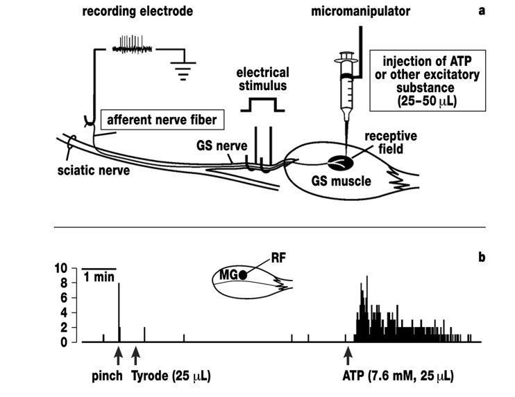
- Reinöhl J, Hoheisel U, Unger T, Mense S. Adenosine triphosphate as a stimulant for nociceptive and non-nociceptive muscle group IV receptors in the rat. Neurosci Lett. 2003;338:25–28. - PubMed
LinkOut - more resources
Full Text Sources
Other Literature Sources
Medical