

Measurements from image-based three dimensional pelvic floor reconstruction: a study of inter- and intraobserver reliability
- PMID: 19629987
- PMCID: PMC2882153
- DOI: 10.1002/jmri.21847
Measurements from image-based three dimensional pelvic floor reconstruction: a study of inter- and intraobserver reliability
Abstract
Purpose: To describe inter- and intraobserver reliability of 3D measurements of female pelvic floor structures.
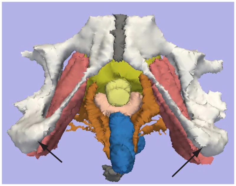
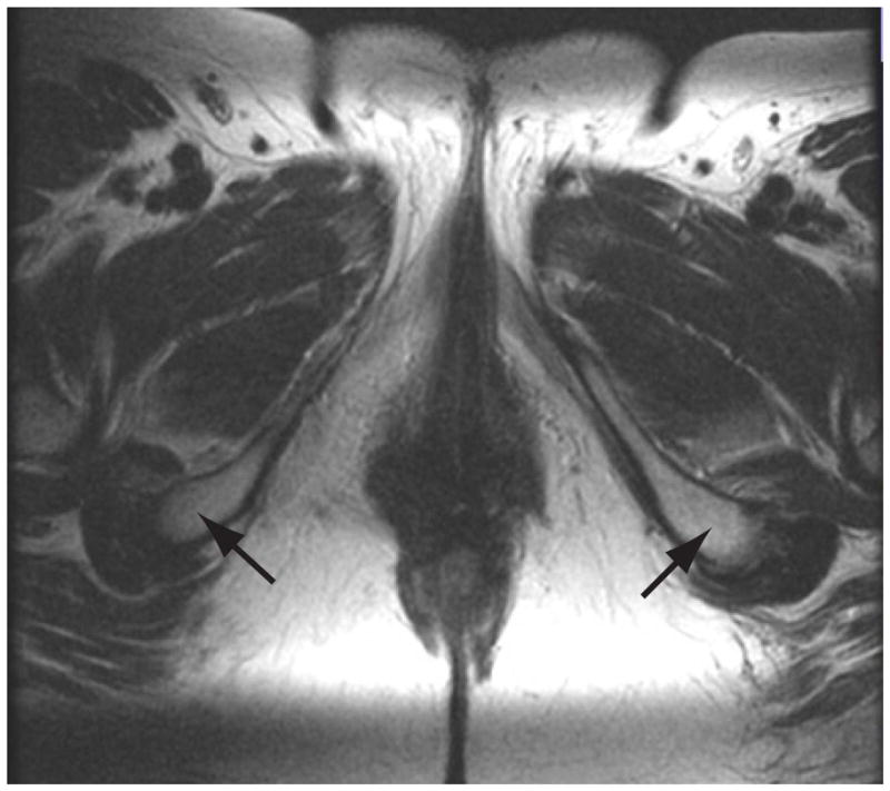
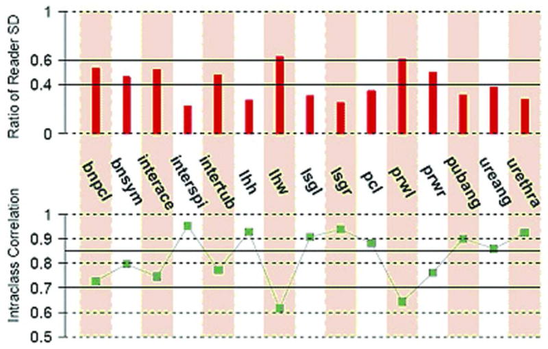
Materials and methods: Twenty reconstructed MR datasets of primiparas at 6-12 months postpartum were analyzed. Pelvic organ measurements were independently made twice by three radiologists blinded to dataset order. A "within-reader" analysis, a "between-reader" analysis, and the intraclass correlation (ICC), and standard deviation ratio (SDR) were computed for each parameter. Fifteen continuous variables and one categorical variable were measured.
Results: Eight continuous parameters showed excellent agreement (ICC >0.85 / SDR <0.40), five parameters showed relatively good agreement (ICC >0.70 / SDR >or=0.40, <0.60). Two parameters showed poor agreement (ICC <or=0.70 and/or SDR >or=0.60). The categorical variable showed poor agreement.
Conclusion: Agreement was best where landmark edges were well defined, acceptable where more "reader judgment" was needed, and poor where levator defects made landmarks difficult to identify. Automated measurement algorithms are under study and may improve agreement in the future.
(c) 2009 Wiley-Liss, Inc.
Figures
References
-
- Fielding JR, Griffiths DJ, Versi E, Mulkern RV, Lee M-LT, Jolesz FA. MR Imaging of Pelvic Floor Continence Mechanisms in the Supine and Upright Positions. Amer J Radiol. 1998;171:1607–1610. - PubMed
-
- Hoyte L, Ratiu P. Linear measurements in 2-dimensional pelvic floor imaging: The impact of slice tilt angles on measurement reproducibility. Am J Obstet Gynecol. 2001;185(3):537–44. - PubMed
-
- Hoyte L, Thomas J, Foster RT, Shott S, Jakab M, Weidner AC. Racial differences in pelvic geometry among asymptomatic nulliparas as seen on three-dimensional MR images. Am J Obstet Gynecol; 2005 Annual Meeting, Soc. Gynecologic Surgeons; California: Rancho Mirage; 2005. - PubMed
-
- Cornella JL, Hibner M, Fenner DE, Kriegshauser JS, Hentz J, Magrina JF. Three-dimensional reconstruction of magnetic resonance images of the anal sphincter and correlation between sphincter volume and pressure. Am J Obstet Gynecol. 2003;189(1):130–5. - PubMed
-
- Singh K, Jakab M, Reid WM, Berger LA, Hoyte L. Three-dimensional magnetic resonance imaging assessment of levator ani morphologic features in different grades of prolapse. Am J Obstet Gynecol. 2003;188(4):910–5. - PubMed
Publication types
MeSH terms
Grants and funding
- U10 HD041261/HD/NICHD NIH HHS/United States
- U01 HD041249/HD/NICHD NIH HHS/United States
- U10 HD41261/HD/NICHD NIH HHS/United States
- U10 HD41248/HD/NICHD NIH HHS/United States
- U10 HD41250/HD/NICHD NIH HHS/United States
- U10 HD41268/HD/NICHD NIH HHS/United States
- U10 HD041263/HD/NICHD NIH HHS/United States
- U10 HD041267/HD/NICHD NIH HHS/United States
- U10 HD041248/HD/NICHD NIH HHS/United States
- U10 HD041269/HD/NICHD NIH HHS/United States
- U10 HD041268/HD/NICHD NIH HHS/United States
- U10 HD041250/HD/NICHD NIH HHS/United States
- U10 HD41267/HD/NICHD NIH HHS/United States
- U10 HD41269/HD/NICHD NIH HHS/United States
- U10 HD41263/HD/NICHD NIH HHS/United States
- U01 HD41249/HD/NICHD NIH HHS/United States
LinkOut - more resources
Full Text Sources
Medical
Miscellaneous
