

Dual-mode intracranial catheter integrating 3D ultrasound imaging and hyperthermia for neuro-oncology: feasibility study
- PMID: 19630251
- PMCID: PMC2810199
- DOI: 10.1177/016173460903100201
Dual-mode intracranial catheter integrating 3D ultrasound imaging and hyperthermia for neuro-oncology: feasibility study
Abstract
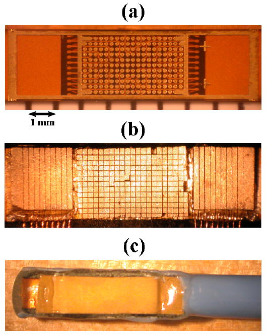
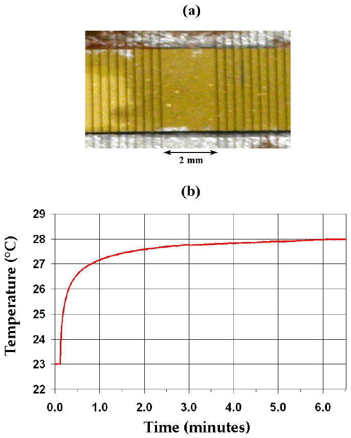
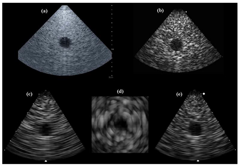
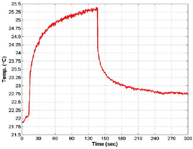
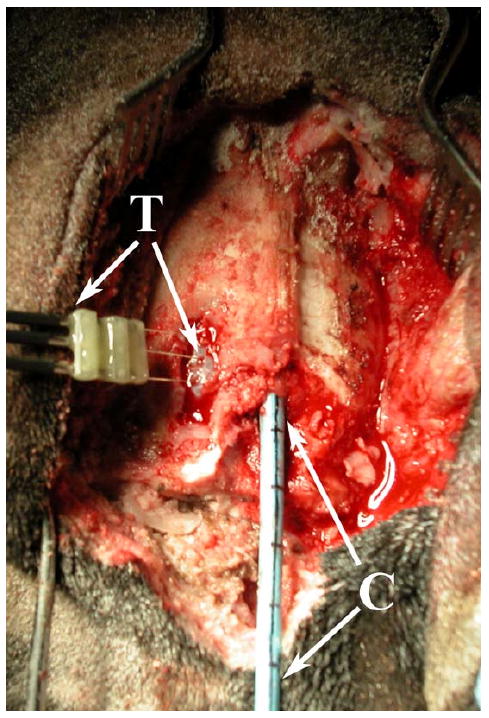
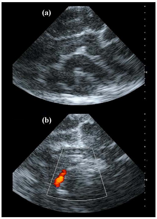
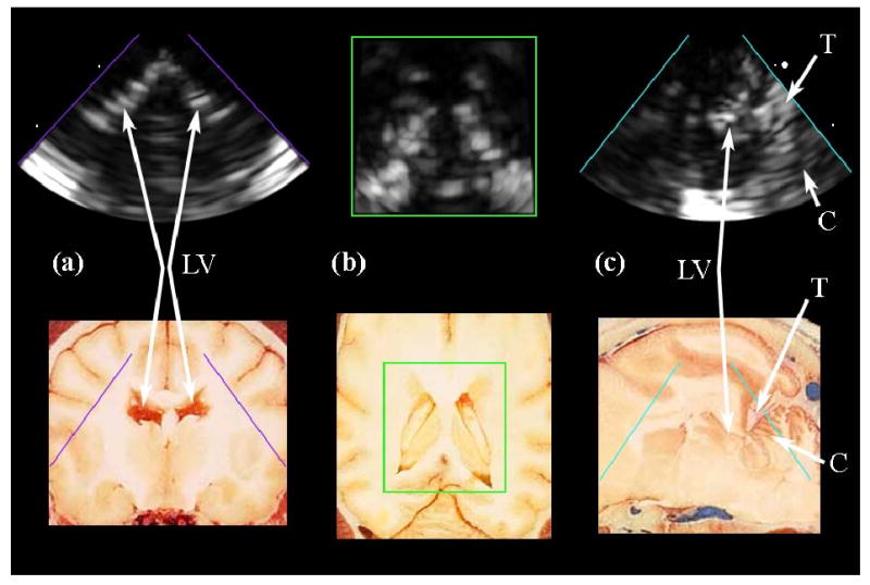
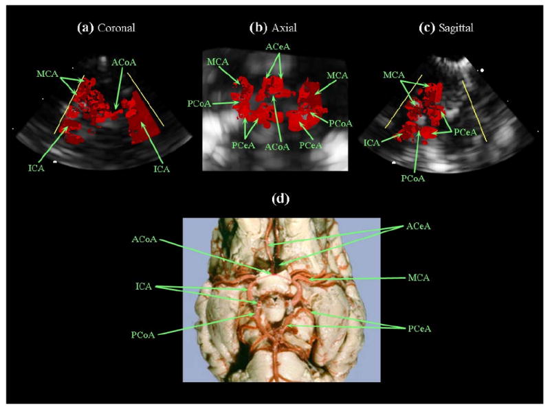
In this study, we investigated the feasibility of an intracranial catheter transducer with dual-mode capability of real-time 3D (RT3D) imaging and ultrasound hyperthermia, for application in the visualization and treatment of tumors in the brain. Feasibility is demonstrated in two ways: first by using a 50-element linear array transducer (17 mm x 3.1 mm aperture) operating at 4.4 MHz with our Volumetrics diagnostic scanner and custom, electrical impedance-matching circuits to achieve a temperature rise over 4 degrees C in excised pork muscle, and second, by designing and constructing a 12 Fr, integrated matrix and linear-array catheter transducer prototype for combined RT3D imaging and heating capability. This dual-mode catheter incorporated 153 matrix array elements and 11 linear array elements diced on a 0.2 mm pitch, with a total aperture size of 8.4 mm x 2.3 mm. This 3.64 MHz array achieved a 3.5 degrees C in vitro temperature rise at a 2 cm focal distance in tissue-mimicking material. The dual-mode catheter prototype was compared with a Siemens 10 Fr AcuNav catheter as a gold standard in experiments assessing image quality and therapeutic potential and both probes were used in an in vivo canine brain model to image anatomical structures and color Doppler blood flow and to attempt in vivo heating.
Figures

Similar articles
-
Intracranial dual-mode IVUS and hyperthermia using circular arrays: preliminary experiments.Ultrason Imaging. 2013 Jan;35(1):17-29. doi: 10.1177/0161734612469372. Ultrason Imaging. 2013. PMID: 23287504 Free PMC article.
-
Integrated endoscope for real-time 3D ultrasound imaging and hyperthermia: feasibility study.Ultrason Imaging. 2007 Jan;29(1):1-14. doi: 10.1177/016173460702900101. Ultrason Imaging. 2007. PMID: 17491295
-
Dual-mode IVUS catheter for intracranial image-guided hyperthermia: feasibility study.IEEE Trans Ultrason Ferroelectr Freq Control. 2010 Nov;57(11):2572-84. doi: 10.1109/TUFFC.2010.1723. IEEE Trans Ultrason Ferroelectr Freq Control. 2010. PMID: 21041144 Free PMC article.
-
Dual-mode IVUS transducer for image-guided brain therapy: preliminary experiments.Ultrasound Med Biol. 2011 Oct;37(10):1667-76. doi: 10.1016/j.ultrasmedbio.2011.06.017. Epub 2011 Aug 19. Ultrasound Med Biol. 2011. PMID: 21856073 Free PMC article.
-
Principles and devices.Semin Vasc Surg. 2006 Sep;19(3):128-31. doi: 10.1053/j.semvascsurg.2006.06.006. Semin Vasc Surg. 2006. PMID: 16996413 Review.
Cited by
-
Intracranial dual-mode IVUS and hyperthermia using circular arrays: preliminary experiments.Ultrason Imaging. 2013 Jan;35(1):17-29. doi: 10.1177/0161734612469372. Ultrason Imaging. 2013. PMID: 23287504 Free PMC article.
-
Ultrasound-based tumor movement compensation during navigated laparoscopic liver interventions.Surg Endosc. 2014 May;28(5):1734-41. doi: 10.1007/s00464-013-3374-9. Epub 2014 Jan 3. Surg Endosc. 2014. PMID: 24385248
-
Advances in diagnostic and treatment modalities for intracranial tumors.J Vet Intern Med. 2014 Jul-Aug;28(4):1165-85. doi: 10.1111/jvim.12370. Epub 2014 May 9. J Vet Intern Med. 2014. PMID: 24814688 Free PMC article. Review.
-
A 9-Fr Endovascular Therapy Transducer With an Acoustic Metamaterial Lens for Rapid Stroke Thrombectomy.IEEE Trans Ultrason Ferroelectr Freq Control. 2024 Nov;71(11):1627-1640. doi: 10.1109/TUFFC.2024.3464330. Epub 2024 Nov 27. IEEE Trans Ultrason Ferroelectr Freq Control. 2024. PMID: 39298303
-
Dual-frequency piezoelectric transducers for contrast enhanced ultrasound imaging.Sensors (Basel). 2014 Nov 4;14(11):20825-42. doi: 10.3390/s141120825. Sensors (Basel). 2014. PMID: 25375755 Free PMC article.
References
-
- ACS. Cancer Facts & Figures 2008. American Cancer Society; 2008.
-
- CBTRUS. Statistical Report: Primary Brain Tumors in the United States, 2000-2004. Central Brain Tumor Registry of the United States; 2008.
-
- Kleihues P, Cavenee WK, International Agency for Research on Cancer., International Society of Neuropathology., International Academy of Pathology., Preuss Foundation for Brain Tumor Research . Pathology and genetics of tumours of the nervous system. IARC Press; 2000.
-
- Mareel M. Anti-Invasive Brain Tumor Therapy: General Aspects and Future Strategies. In: Mikkelsen T, editor. Brain Tumor Invasion: Biological, Clinical, and Therapeutic Considerations. Wiley-Liss; 1998.
-
- Prados MD. Systemic Chemotherapy. In: Bernstein M, Berger MS, editors. Neuro-oncology: The Essentials. Thieme Medical Publishers; 2000.
Publication types
MeSH terms
Grants and funding
LinkOut - more resources
Full Text Sources
Medical
