. 2009 Sep 1;48(34):8241-8.
doi: 10.1021/bi9007879.

Calcium-dependent lateral organization in phosphatidylinositol 4,5-bisphosphate (PIP2)- and cholesterol-containing monolayers

Affiliations

Calcium-dependent lateral organization in phosphatidylinositol 4,5-bisphosphate (PIP2)- and cholesterol-containing monolayers

Ilya Levental et al. Biochemistry. .

Abstract

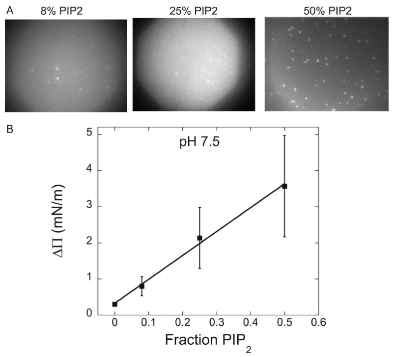
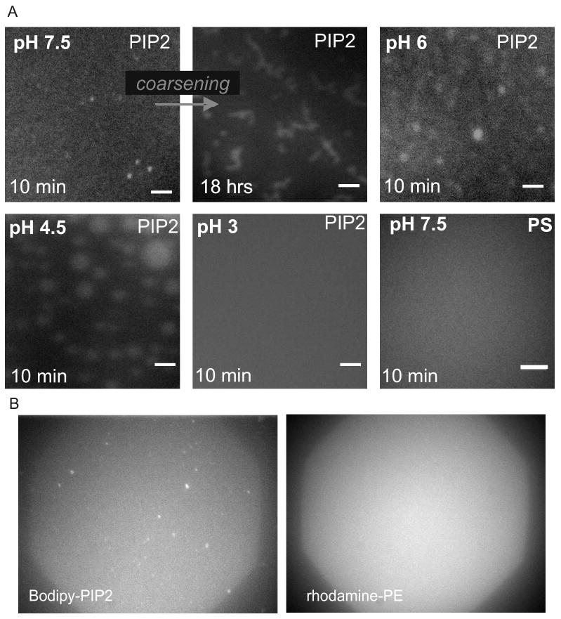
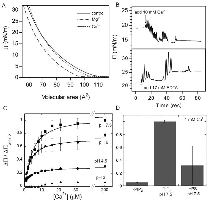
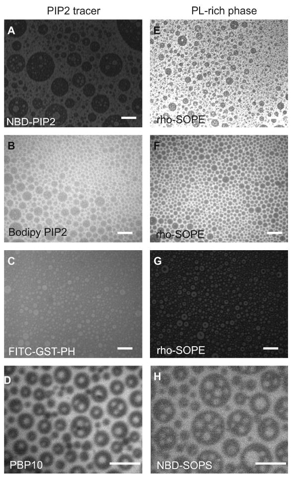
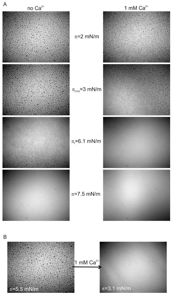
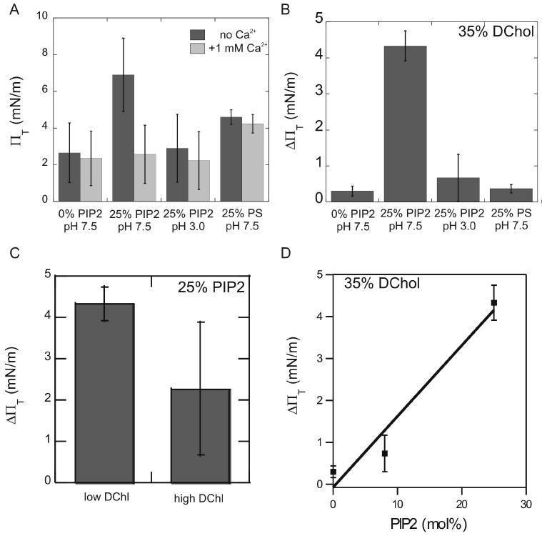
Biological membrane function, in part, depends upon the local regulation of lipid composition. The spatial heterogeneity of membrane lipids has been extensively explored in the context of cholesterol and phospholipid acyl-chain-dependent domain formation, but the effects of lipid head groups and soluble factors in lateral lipid organization are less clear. In this contribution, the effects of divalent calcium ions on domain formation in monolayers containing phosphatidylinositol 4,5-bisphosphate (PIP2), a polyanionic, multifunctional lipid of the cytosolic leaflet of the plasma bilayer, are reported. In binary monolayers of PIP2 mixed with zwitterionic lipids, calcium induced a rapid, PIP2-dependent surface pressure drop, with the concomitant formation of laterally segregated, PIP2-rich domains. The effect was dependent upon head-group multivalency, because lowered pH suppressed the surface-pressure effect and domain formation. In accordance with previous observations, inclusion of cholesterol in lipid mixtures induced coexistence of two liquid phases. Phase separation strongly segregated PIP2 to the cholesterol-poor phase, suggesting a role for cholesterol-dependent lipid demixing in regulating PIP2 localization and local concentration. Similar to binary mixtures, subphase calcium induced contraction of ternary cholesterol-containing monolayers; however, in these mixtures, calcium induced an unexpected, PIP2- and multivalency-dependent decrease in the miscibility phase transition surface pressure, resulting in rapid dissolution of the domains. This result emphasizes the likely critical role of subphase factors and lipid head-group specificity in the formation and stability of cholesterol-dependent domains in cellular plasma membranes.


Figures

Figure 1
Figure 1. Calcium-induced change in surface pressure and domain formation linearly depends on PIP2 fraction
(A) The formation of calcium-induced PIP2 domains (visualized by the inclusion of 0.5% Bodipy-PIP2) is persistent from 8 to 50 mol % PIP2, with the abundance of bright domains directly related to the PIP2 fraction. While the change in ∏ is rapid for all compositions (faster than mixing), the time required for PIP2 domains to become microscopically visible at 8% PIP2 is significantly longer than at 25 or 50% and likely reflects diffusion limitations. (B) Decrease in ∏ upon the addition of 1 mM Ca2+ is proportional (R2 = 0.995) to the fraction of PIP2 in the lipid monolayer (balance lipid is SOPC).
Figure 2
Figure 2. Divalent calcium induces microscopic PIP2 domains in mixed lipid monolayers
(A) Fluorescence micrographs of SOPC:PIP2 (3:1) monolayers with 1% NBD-PIP2 or SOPC:SOPS (3:1) with 1% NBD-PS on buffered subphases at varying pH. Addition of 1 mM Ca2+ to the subphase induced microscopically-visible PIP2 domains within minutes at conditions where PIP2 is multivalent (pH 7.5, 6, and 4.5), whereas no monolayer heterogeneity was observed at conditions under which only monovalent and zwitterionic lipids were present (PIP2 at pH 3 and PS at pH 7.5). Although formation of domains was rapid (< 10 mins) PIP2 domains at pH 7.5 coarsened to form micron-sized ribbon-like domains after many hours. Scale bars are 10 μm. All charged lipids added at 25 mol%. (B) To ensure that the domains observed were PIP2-enriched and not membrane defects, monolayers were also formed with 0.1% of the membrane marker, rho-PE. At conditions that result in the formation of PIP2 domains (B-left), epifluorescence micrographs of rho-PE show the monolayer to be homogeneous (B-right) and thus free of artifacts resulting from fluorescent lipids or monolayer collapse.
Figure 3
Figure 3. Subphase calcium induces immediate, reversible, pH-dependent surface pressure decrease in PIP2 containing monolayers
(A) ∏-molecular area isotherms of SOPC:PIP2 (3:1) monolayers show reduced surface pressures in the presence of divalent cations, with greater reductions with Ca2+ compared to Mg2+. (B) The surface pressure (∏) of these monolayers decreases quickly and substantially upon addition of CaCl2 to the pH 7.5 buffered subphase. The time scale (∼10 secs) for the pressure change reflects introduction and mixing of the subphase and is therefore an upper limit on the-time required for Ca2+-mediated contraction (A, top). Addition of excess EDTA to the subphase results in an immediate increase in surface pressure equal in magnitude to the calcium-induced decrease (A, bottom). (C) The change in surface pressure (Δ∏ normalized to Δ∏ with 1 mM Ca2+ at pH 7.5) of a SOPC:PIP2 monolayer as a function of Ca2+ concentration can be modeled by first-order reversible binding with dissociation constant (Kd) independent of subphase pH. Error bars at pH 7.5 and 6 show the typical standard deviation of three separate experiments. (D) Quantification of the total decrease in monolayer surface pressure after 1 mM Ca2+ addition for monolayers of varying composition and subphase pH. Monolayers containing charged lipids show a significant decrease in surface pressure in the presence of divalent cations that is strongly dependent on total surface charge.
Figure 4
Figure 4. PIP2 co-localizes with PL-rich phase markers in fluid-fluid coexistence monolayers
Monolayers of PIP2, cholesterol, and SOPC co-stained with PIP2 markers (A-D) against markers of the PL-rich phase (E-H). Colocalization of PIP2 and markers indicates the exclusion of PIP2 from cholesterol-rich domains at ∏ below ∏T. Compositions were – (A/E) DChol:SOPC:PIP2 (50:40:10) + 1% NBD-PIP2 + 0.1% rho-SOPE; (B/F) DChol:SOPC:PIP2 (50:40:10) + 0.5% BodipyFL-PIP2 + 0.1% rho-SOPE; (C/G) DChol:SOPC:PIP2 (50:40:10) + 0.1% rho-SOPE; and (D/H) DChol:SOPC:PIP2 (50:40:10) + 1% NBD-PC. Scale bars are 10 μm.
Figure 5
Figure 5. Subphase calcium decreases monolayer ∏T in cholesterol containing monolayers
(A) Fluorescence micrographs of SOPC:PIP2:DChol (40:25:35) monolayers, which form PL-rich (bright) and Chol-rich (dark) domains at ∏< ∏T (visualized by the inclusion of 0.1% Rho-SOPE). As ∏ is increased through ∏T, the monolayer becomes mixed and homogeneously fluorescent. The addition of 1 mM Ca2+ to the subphase results in a decrease in ∏T from ∏T = 6.1 mN/m to ∏T,Ca = 3.0 mN/m as shown in the two series of panels. (B) The effect of calcium on ∏T was tested on demixed monolayers where ∏ < ∏T. Ca2+ (1 mM) added to the subphase resulted in a rapid decrease in ∏ and mixing of the monolayer as ∏ > ∏T,Ca.
Figure 6
Figure 6. Presence of subphase Ca2+ affects ∏T only in monolayers containing highly charged lipids
(A)T for 35% cholesterol monolayers increases significantly (p=0.046) with the incorporation of PIP2 at pH 7.5 in the absence of Ca2+ (dark bars); the effect is counteracted by the presence of 1 mM Ca2+ (light gray bars). This increase is dependent on the multivalent nature of PIP2 (p<0.001), as no significant increase in ∏T was observed with PIP2 at pH 3 (where the net charge of PIP2 is approximately -1; p=0.5) or with incorporation of PS (net charge -1; p=0.6). (B) The calcium-induced decrease of ∏T (Δ∏T = ∏T - ∏T,Ca) depends strongly on the multivalency of PIP2 at pH 7.5 where Δ∏T = 4.3 ± 0.4 mN/m. For monolayers with only net neutral lipids, or those with monoanionic lipids (PIP2 at pH 3, PS at pH 7.5), the addition of Ca2+ induced only a small Δ∏T. (C) For monolayers of constant PIP2 fraction, the effect of calcium addition on ∏T is weakly dependent on the amount of cholesterol (p=0.1). (D) For monolayers of 35% DChol, Δ∏T upon addition of calcium depends linearly (R2 = 0.952) on the amount of PIP2 incorporated into the monolayer. All error bars shown are standard deviations from 3 separate experiments. All lipid mixtures included 0.1% rho-SOPE for fluorescent visualization, and the balance of the lipid mixture was SOPC. All charged lipids were included at 25 mol% unless otherwise noted.

Similar articles

Cited by

References

    1. Esposti MD. Lipids, cardiolipin and apoptosis: a greasy license to kill. Cell Death Differ. 2002;9:234–236. - PubMed
    1. Lawrence T, Willoughby DA, Gilroy DW. Anti-inflammatory lipid mediators and insights into the resolution of inflammation. Nat Rev Immunol. 2002;2:787–795. - PubMed
    1. Yin HL, Janmey PA. Phosphoinositide regulation of the actin cytoskeleton. Annu Rev Physiol. 2003;65:761–789. - PubMed
    1. Eling TE, Glasgow WC. Cellular proliferation and lipid metabolism: importance of lipoxygenases in modulating epidermal growth factor-dependent mitogenesis. Cancer Metastasis Rev. 1994;13:397–410. - PubMed
    1. Czech MP. PIP2 and PIP3: complex roles at the cell surface. Cell. 2000;100:603–606. - PubMed

Publication types