

MRI-guided focused ultrasound surgery
- PMID: 19630579
- PMCID: PMC4005559
- DOI: 10.1146/annurev.med.60.041707.170303
MRI-guided focused ultrasound surgery
Abstract
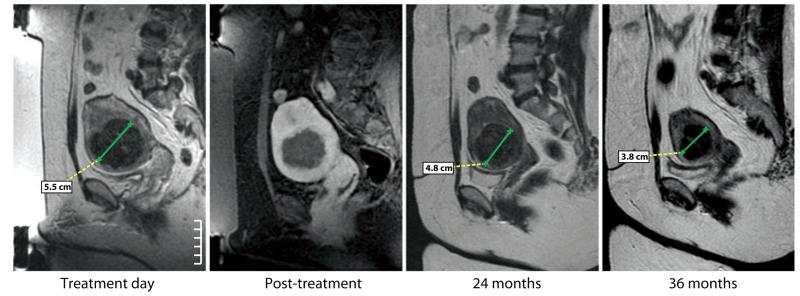
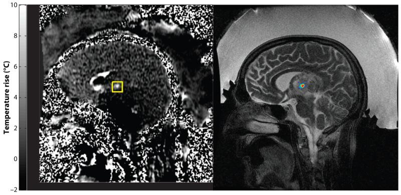
MRI-guided focused ultrasound (MRgFUS) surgery is a noninvasive thermal ablation method that uses magnetic resonance imaging (MRI) for target definition, treatment planning, and closed-loop control of energy deposition. Integrating FUS and MRI as a therapy delivery system allows us to localize, target, and monitor in real time, and thus to ablate targeted tissue without damaging normal structures. This precision makes MRgFUS an attractive alternative to surgical resection or radiation therapy of benign and malignant tumors. Already approved for the treatment of uterine fibroids, MRgFUS is in ongoing clinical trials for the treatment of breast, liver, prostate, and brain cancer and for the palliation of pain in bone metastasis. In addition to thermal ablation, FUS, with or without the use of microbubbles, can temporarily change vascular or cell membrane permeability and release or activate various compounds for targeted drug delivery or gene therapy. A disruptive technology, MRgFUS provides new therapeutic approaches and may cause major changes in patient management and several medical disciplines.
Figures
References
-
- Haemmerich D, Laeseke PF. Thermal tumour ablation: devices, clinical applications and future directions. Int. J. Hyperthermia. 2005;21(8):755–60. - PubMed
-
- Jolesz FA. Image guidance and control of thermal ablation. In: van Sonnenberg E, McMullen W, Solbiati L, editors. Tumor Ablation: Principles and Practice. Vol. 1. Springer-Verlag; New York: 2005. pp. 182–91.
-
- Jolesz FA, Blumenfeld SM. Interventional use of magnetic resonance imaging. Magn. Reson. Q. 1994;10(2):85–96. - PubMed
-
- Black PM, Moriarty T, Alexander E, 3rd, et al. Development and implementation of intraoperative magnetic resonance imaging and its neurosurgical applications. Neurosurgery. 1997;41(4):831–42. discussion 842-45. - PubMed
-
- Nimsky C, Ganslandt O, Kober H, et al. Intraoperative magnetic resonance imaging combined with neuronavigation: a new concept. Neurosurgery. 2001;48(5):1082–89. - PubMed
Publication types
MeSH terms
Grants and funding
LinkOut - more resources
Full Text Sources
Other Literature Sources
Medical
