. 2009 Nov 15;394(2):171-6.
doi: 10.1016/j.ab.2009.07.027. Epub 2009 Jul 22.

An adhesion-based method for plasma membrane isolation: evaluating cholesterol extraction from cells and their membranes

Affiliations

An adhesion-based method for plasma membrane isolation: evaluating cholesterol extraction from cells and their membranes

Ludmila Bezrukov et al. Anal Biochem. .

Abstract

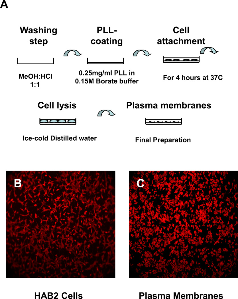
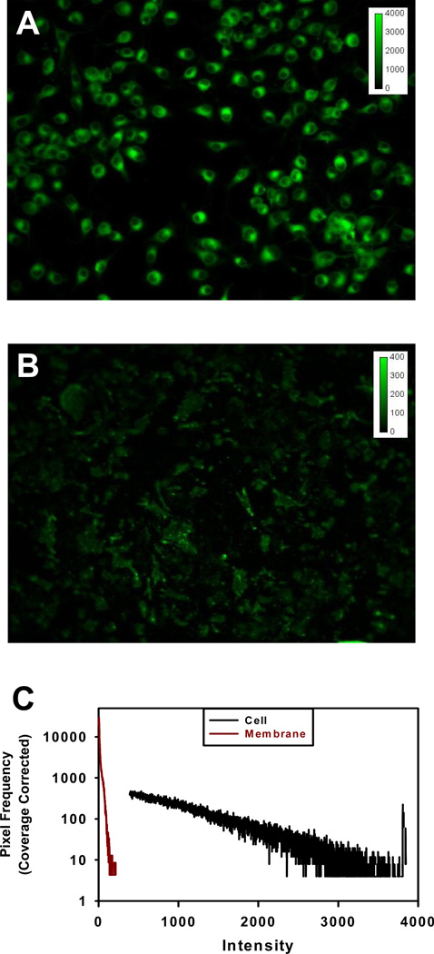
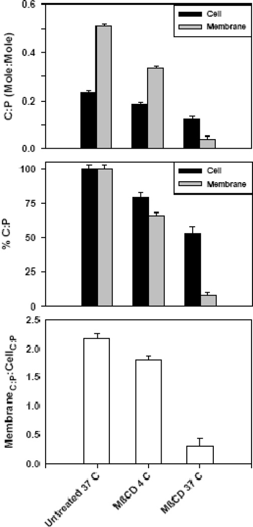
A method to isolate large quantities of directly accessible plasma membrane from attached cells is presented. The method is based on the adhesion of cells to an adsorbed layer of polylysine on glass plates, followed by hypotonic lysis with ice-cold distilled water and subsequent washing steps. Optimal conditions for coating glass plates and time for cell attachment were established. No additional chemical or mechanical treatments were used. Contamination of the isolated plasma membrane by cell organelles was less than 5%. The method uses inexpensive, commercially available polylysine and reusable glass plates. Plasma membrane preparations can be made in 15 min. Using this method, we determined that methyl-beta-cyclodextrin differentially extracts cholesterol from fibroblast cells and their plasma membranes and that these differences are temperature dependent. Determination of the cholesterol/phospholipid ratio from intact cells does not reflect methyl-beta-cyclodextrin plasma membrane extraction properties.


Figures

Figure 1
Figure 1. Plasma membrane isolation
A. Glass plates were soaked in Hydrochloric Acid: Methanol (1:1) at room temperature, washed with water, rinsed with ethanol, and dried; glass plates were coated with polylysine; cells were incubated at 37 °C for 4 hours; cells were ruptured with ice-cold distilled water; intracellular debris was removed with ice-cold TBS buffer washes. B. HAB2 cells on a polylysine coated glass plate after incubation for 4 hours at 37 °C in serum-free media. C. Plasma membranes of HAB2 cells on a polylysine coated glass plate. Both samples were stained with 1µM PKH 26 for 10 min at room temperature.
Figure 2
Figure 2. Staining of Golgi apparatus and purity of plasma membrane preparation
HAB2 cells (A) and plasma membranes of HAB2 cells (B) were stained with 5 µM BODYPY FL C5-ceramide (green) specific for Golgi apparatus for 10 min at 37 °C in serum-free medium (DMEM) and visualized using a 32× objective. (C) Intensity histogram used to calculate membrane purity. In this example, the Signal Area, ifi*Ii for the HAB2 cells and plasma membranes were 8.79*107 and 3.59*106 and the number of background pixels, 2.39*105 and 2.50*105respectively. With 3.22*105 total pixels, the coverage correction for cells and membranes is, 0.26 and 0.22. The corresponding Signalcorrectedare 3.41*108 and 1.60*107 representing a purity of 4.7%. For direct comparison, the pixel frequency, fi, was normalized by the coverage correction, fi/0.26 and fi/0.22, respectively, and designated as Coverage Corrected on the axis.
Figure 3
Figure 3. Cholesterol:phospholipid (C:P) at different extraction temperatures
(A) C:P ratio for untreated and MβCD extracted HAB2 cells and their plasma membranes. Data are averages of 6 – 11 experiments. (B) Normalized data from Fig. 4-A, where 100% is the ratio of cholesterol to phospholipids in untreated (control) samples. C) Membrane C:P to Cell C:P, a measure of the relative concentration of cholesterol in the plasma membranes compared to intact cells.

Similar articles

Cited by

References

    1. Depierre JW, Karnovsky ML. Plasma membranes of mammalian cells. J. Cell. Biol. 1973;56:275–303. - PMC - PubMed
    1. Atkinson PH, Summers DF. Purification and properties of HeLa cell plasma membranes. J. Biol. Chem. 1971;246:5162–5175. - PubMed
    1. Boone CW, Ford LE, Stuart DC, Lorenz D D. Isolation of plasma membrane fragments from Hela cells. J. Cell. Biol. 1969;41:378–392. - PMC - PubMed
    1. Bosmann HB, Hagopian A, Eylar EH. Cellular membranes: the isolation and characterization of the plasma and smooth membranes of HeLa cells. Arch. Biochem. Biophys. 1968;128:51–59. - PubMed
    1. Johnsen S, Stokke T, Prydz H. HeLa cell plasma membranes. I.5-nucleotidase and ouabain-sensetive ATPase as markers for plasma membranes. J. Cell. Biol. 1974;63:357–363. - PMC - PubMed

Publication types

MeSH terms