. 2009 Oct 1;334(1):174-85.
doi: 10.1016/j.ydbio.2009.07.015. Epub 2009 Jul 22.

Wnt/beta-catenin signaling plays an essential role in activation of odontogenic mesenchyme during early tooth development

Affiliations

Wnt/beta-catenin signaling plays an essential role in activation of odontogenic mesenchyme during early tooth development

Jianquan Chen et al. Dev Biol. .

Abstract

Classical tissue recombination studies demonstrated that initiation of tooth development depends on activation of odontogenic potential in the mesenchyme by signals from the presumptive dental epithelium. Although several members of the Wnt family of signaling molecules are expressed in the presumptive dental epithelium at the beginning of tooth initiation, whether Wnt signaling is directly involved in the activation of the odontogenic mesenchyme has not been characterized. In this report, we show that tissue-specific inactivation of beta-catenin, a central component of the canonical Wnt signaling pathway, in the developing tooth mesenchyme caused tooth developmental arrest at the bud stage in mice. We show that mesenchymal beta-catenin function is required for expression of Lef1 and Fgf3 in the developing tooth mesenchyme and for induction of primary enamel knot in the developing tooth epithelium. Expression of Msx1 and Pax9, two essential tooth mesenchyme transcription factors downstream of Bmp and Fgf signaling, respectively, were not altered in the absence of beta-catenin in the tooth mesenchyme. Moreover, we found that constitutive stabilization of beta-catenin in the developing palatal mesenchyme induced aberrant palatal epithelial invaginations that resembled early tooth buds both morphologically and in epithelial molecular marker expression, but without activating expression of Msx1 and Pax9 in the mesenchyme. Together, these results indicate that activation of the mesenchymal odontogenic program during early tooth development requires concerted actions of Bmp, Fgf and Wnt signaling from the presumptive dental epithelium to the mesenchyme.


Figures
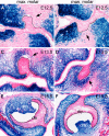
Fig. 1
Fig. 1
Cre-mediated activation of LacZ expression during molar tooth development in the Osr2-IresCre; R26R embryos. (A-F) Frontal sections from Osr2-IresCre; R26R embryos at E12.5 (A, B), E13.5 (C, D), and E15.5 (E, F) were assayed by X-gal staining. Note that X-gal staining was only detected in dental mesenchymal cells, but not in dental epithelia. Black dashed lines mark the boundary between the developing dental epithelium and mesenchyme. Arrows in A-D point to the mesenchyme lingual to the developing tooth buds. de, dental epithelium; dm, dental mesenchyme; max. molar, maxillary first molar tooth germs; man. molar, mandibular first molar tooth germs.
Fig. 2
Fig. 2
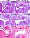
Molar tooth development arrested at the bud stage in the Catnbf/f; Osr2-IresCre mutant mice. (A-F) Histology of frontal sections through the developing molar tooth germs in control (A, C, E) and Catnbf/f; Osr2-IresCre mutant (B, D, F) embryos at E13.5 (A, B), E14.5 (C, D) and E15.5 (E, F). Arrows point to the molar tooth germs.
Fig. 3
Fig. 3
Incisor tooth development arrested at the late bud to early cap stages in the Catnbf/f; Osr2-IresCre mutant mice. White arrows point to mandibular tooth germs, and black arrows point to maxillary tooth germs. (A-D) At E13.5, both maxillary and mandibular incisors developed to the bud stage in control (A, C) and Catnbf/f; Osr2-IresCre mutant (B, D) embryos. (E-H) At E14.5, the incisor tooth germs have developed to the cap stage in the control embryos (E, G) but incisor tooth germs in the Catnbf/f; Osr2-IresCre mutant littermates (F, H) were still at the bud stage. (I-L) At E15.5, the incisor tooth germs developed to bell stage in the control embryos (I, K) but the mandibular incisors arrested at the bud stage whereas the maxillary incisor tooth germs arrested at the early cap stage in the Catnbf/f; Osr2-IresCre mutant embryos. Ectopic cartilage (ec) developed next to the incisor tooth germs in the Catnbf/f; Osr2-IresCre mutant embryos by E15.5 (J).
Fig. 4
Fig. 4
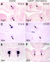
β-catenin function is required in the developing tooth mesenchyme for induction of primary enamel knot. (A) At E14.5, Fgf4 mRNA was strongly expressed in the primary enamel knot cells (arrows) in the developing first molar tooth germs in the control embryo. (B) Fgf4 expression was not detected in the developing first molar tooth germs in the E14.5 Catnbf/f; Osr2-IresCre mutant embryos. Arrows point to the developing tooth germs. (C) At E14.5, Shh mRNA was strongly expressed in the primary enamel knot cells in the developing first molar tooth germs in the control embryo. (D) Shh mRNA expression was detected in a few cells in the maxillary first molar tooth germ but not in the mandibular molar tooth germ in the E14.5 Catnbf/f; Osr2-IresCre mutant embryo. (E, F) At E14.5, Shh mRNA was strongly expressed in the inner enamel epithelium in the developing incisors in control embryo (E) but was only weakly expressed in a few cells in the developing incisor epithelium in the Catnbf/f; Osr2-IresCre mutant littermate (F).
Fig. 5
Fig. 5
Tooth developmental arrest in the Catnbf/f; Osr2-IresCre mutant mice was not accompanied by increased cell apoptosis. (A, C) At E14.5, the primary enamel knot cells at the center of the developing inner enamel epithelium in the maxillary (A) and mandibular (C) molar tooth germs in the control embryo exhibited active caspase-3 activity. (B, D) A rudimentary primary enamel knot, as shown by a few clustered cells expressing active caspase-3, formed in the developing maxillary molar tooth germ (B), but not in the mandibular molar tooth germ (D), in the E14.5 Catnbf/f; Osr2-IresCre mutant embryos. Arrows in A – D point to the corresponding primary enamel knot region of the developing molar tooth germs. No active caspase-3 activity was detected in the developing tooth mesenchyme in either the control (A, C) or Catnbf/f; Osr2-IresCre mutant (B, D) embryos. (E-H) Detection of active caspase-3 activity in the developing molar tooth germs in E12.5 (E, F) and E13.5 (G, H) control (E, G) and Catnbf/f; Osr2-IresCre mutant (F, H) embryos. Arrowheads in E – H point to the developing tooth buds. No differences in cell apoptosis were detected in the control and mutant tooth germs.
Fig. 6
Fig. 6
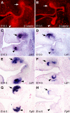
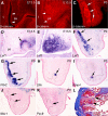
Alterations in gene expression during tooth development in the Catnbf/f; Osr2-IresCre mutant embryos. (A, B) Immunofluorescent detection of β-catenin protein expression in the developing molar tooth germs in E13.5 control (A) and Catnbf/f; Osr2-IresCre mutant (B) littermates. Arrows point to developing maxillary molar tooth mesenchyme and arrowheads point to the developing mandibular molar tooth mesenchyme. (C-F) Lef1 mRNA expression in E13.5 (C, D) and E14.5 (E, F) control (C, E) and Catnbf/f; Osr2-IresCre mutant (D, F) molar tooth germs, respectively. (G, H) Fgf3 mRNA expression was dramatically reduced in the E14.5 Catnbf/f; Osr2-IresCre mutant molar tooth germs (H), in comparison with that in the control littermate (G).
Fig. 7
Fig. 7
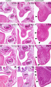
Constitutive stabilization of β-catenin in the developing palatal mesenchyme resulted in ectopic tooth-like epithelial buds in the palate. (A, B) At E15.5, the Catnblox(ex3)/+;Osr2-CreKI mutant embryo (B) exhibited comparable molar tooth development with that in the control littermate (A), but the mutant palatal shelves were still vertically oriented (B) while palatal shelves had elevated to the horizontal position above the tongue in the control littermates. (C) High magnification view of the Catnblox(ex3)/+;Osr2-CreKI mutant palatal shelf at E15.5. Arrowhead points to thickened palatal epithelium. (D - F) At E16.5, while the developing molar tooth germs exhibited similar morphology in control (D) and the Catnblox(ex3)/+;Osr2-CreKI mutant (E) littermates, the mutant palatal shelves (F) exhibited epithelial invaginations (arrowheads in F). (G-I) At E17.5, the molar tooth germs appear smaller in the Catnblox(ex3)/+;Osr2-CreKI mutant (H) than those in the control embryo (G). Some of the epithelial invaginations in the mutant palate (I) appeared to form early “cap”-like structures (arrowheads in I). (J-L) At P0, the molar tooth germs in the Catnblox(ex3)/+;Osr2-CreKI mutant (K) appeared retarded, in comparison with those in control littermate (J). Multiple epithelial invaginations (arrowheads in L) were detected in the mutant palatal shelves and the palatal mesenchyme appeared to condense under the epithelial invaginations.
Fig. 8
Fig. 8
Constitutive stabilization of β-catenin in the palatal mesenchyme induced tooth bud-like structures from the palatal epithelium. (A-C) Immunofluorescent detection of β-catenin protein in the palate in E13.5 (A, B) and P0 (C) control (A) and Catnblox(ex3)/+;Osr2-CreKI mutant (B, C) mice. Much more β-catenin protein (red color) accumulated in the developing palate mesenchyme in E13.5 Catnblox(ex3)/+;Osr2-CreKI mutant (B) than in the control littermate (A). By P0, β-catenin protein was preferentially accumulated in the mesenchymal cells underlying the epithelial invaginations in the mutant palate (C). Arrowheads in A and B point to the palatal epithelium, whereas arrows in C point to the β-catenin positive palatal mesenchyme underlying the invaginated palatal epithelium. (D-F) In situ hybridization detection of Lef1 mRNA expression (blue color) in the palate in E13.5 (D, E) and P0 (F) control (D) and Catnblox(ex3)/+;Osr2-CreKI mutant (E, F) mice. Arrow in D points to Lef1-positive tooth mesenchyme, whereas arrows in F point to Lef1 mRNA expression in the palatal mesenchyme underlying the invaginated palatal epithelium in the Catnblox(ex3)/+;Osr2-CreKI mutant mouse. (G-I) Expression of Pitx2 (G), Shh (H), and Bmp4 (I) mRNAs indicate that the invaginated epithelial structures in the palate in the Catnblox(ex3)/+;Osr2-CreKI mutant mice resemble developing tooth germs. Arrows in G point to Pitx2 mRNA expression in the invaginated palatal epithelium. Arrows in H and I point to the Shh- and Bmp4-positive domain in the distal region of the invaginated palatal epithelium, which resemble primary enamel knot in gene expression pattern. (J, K) The mesenchyme underlying palatal epithelial invaginations (arrows) in Catnblox(ex3)/+;Osr2-CreKI mutant mice did not show detectable expression of Msx1 (J) and Pax9 mRNA (K). Black dashed line in each panel mark the boundary between the invaginated epithelium and mesenchyme. (L) Kidney capsule graft of E13.5 Catnblox(ex3)/+;Osr2-CreKI mutant palatal shelf had encapsulated cysts (arrows) but no organized tooth structures.
Fig. 9
Fig. 9
Persistent stabilization of β-catenin in the tooth mesenchyme disrupts cytodifferentiation during later tooth morphogenesis in the Catnblox(ex3)/+;Osr2-CreKI mutant mice. (A, B) Frontal sections through the mandibular first molar tooth germs of control (A) and Catnblox(ex3)/+;Osr2-CreKI mutant mice at P0 stained with hematoxylin and eosin. Note the uniform distribution of dental mesenchymal cells in the dental pulp in the control mouse versus the disorganized dental pulp compartment in the mutant mouse. (C, D) Immunostaining showing β-catenin distribution (red color) in the control (C) and Catnblox(ex3)/+;Osr2-CreKI mutant (D) molar tooth germs at P0. Cellular nuclei were counterstained with 4’-6-diamidino-2-phenylindole (DAPI) and shown in blue color. Note the much higher level of β-catenin protein in the dental mesenchyme of the mutant mouse (D) than that in the control littermate (C). (E, F) In situ hybridization detection of Bmp4 mRNA (shown in blue color) in the frontal sections of molar tooth germs in control (E) and Catnblox(ex3)/+;Osr2-CreKI mutant (F) mice. (G, H) Immunohistochemical staining (detected in brown color) of the frontal sections of molar tooth germs in control (G) and Catnblox(ex3)/+;Osr2-CreKI mutant (H) mice with an antibody against phosporylated-Smad1/5/8. The sections were counterstained with hematoxylin (blue). (I, J) In situ hybridization detection of Shh mRNA expression in the molar tooth germs in control (I) and Catnblox(ex3)/+;Osr2-CreKI mutant (J) mice. (K, L) Kidney capsule grafts of the molar tooth germs from E13.5 control (K) and Catnblox(ex3)/+;Osr2-CreKI mutant (L) embryos assayed by trichrome staining. The enamel matrix (arrows) is stained red and the dentin matrix is stained blue. Dp, dental pulp.

Similar articles

Cited by

References

    1. Andl T, Reddy ST, Gaddapara T, Millar SE. WNT signals are required for the initiation of hair follicle development. Dev. Cell. 2002;2:643–653. - PubMed
    1. Andl T, Ahn K, Kairo A, Chu EY, Wine-Lee L, Reddy ST, Croft NJ, Cebra-Thomas JA, Metzger D, Chambon P, Lyons KM, Mishina Y, Seykora JT, Crenshaw EB, III, Millar SE. Epithelial Bmpr1a regulates differentiation and proliferation in postnatal hair follicles and is essential for tooth development. Development. 2004;131:2257–2268. - PubMed
    1. Barolo S. Transgenic Wnt/TCF pathway reporters: all you need is Lef? Oncogene. 2006;25:7505–7511. - PubMed
    1. Bei M, Maas R. FGFs and BMP4 induce both Msx1-independent and Msx1-dependent signaling pathways in early tooth development. Development. 1998;125:4325–4333. - PubMed
    1. Bei M, Kratochwil K, Maas RL. BMP4 rescues a non-cell-autonomous function of Msx1 in tooth development. Development. 2000;127:4711–4718. - PubMed

Publication types

MeSH terms

LinkOut - more resources